By Paul T. Finger, MD
History
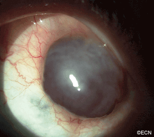
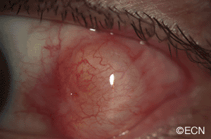
Two subconjunctival masses appearing 45 (Case 1) and 7 (Case 2) years after surgery.
Impression
Giant Conjuctival Inclusion Cysts
Comment
Conjunctival cysts typically cause cosmetic defects and Case 1, prevented proper eyelid closure. Since most are excised, high frequency ultrasound can help define the tumors extent. In Case 2, it differentiated between a solid and cystic pigmented conjunctival tumor.
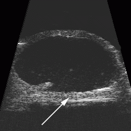
Case 1: High-frequency ultrasound revealed that the lateral rectus muscle was not incarcerated within the tumor and defined the posterior tumor margin. The sclera was found to be intact. This was expected, but not known by slit-lamp examination. These findings were helpful for pre-operative planning.
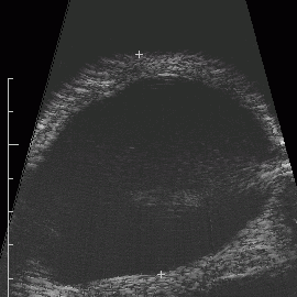
Case 2: This tumor was more ominous looking. It was cystic, pigmented and close to the lacrimal gland. Again, high-frequency ultrasound was used to delineate the tumor’s edges, confirm scleral integrity, and that it was a cyst.
These cases were presented to aid in your continued care of patients with conjunctival cysts. Next time you plan excision of a large conjunctival cyst, you may want to consider evaluating it with a high-frequency ultrasound.