By Paul T. Finger, MD
History

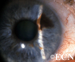
A 79-year-old female with a past history of hypertension and emphysema was referred for preoperative evaluation of a pigmented iris tumor in an eye with a 20/80 cataract OS.
At the The New York Eye Cancer Center her iris tumor was noted to exhibit ectropion uveae, pigment liberation into the angle, and sector cataract. Intraocular pressure measurements were 17 OU.
Indications for removal of presumed iris melanomas include: growth and secondary glaucoma. In this case there was neither. There is some controversy as to whether to remove or biopsy such lesions at the time of cataract surgery. After a detailed discussion of the potential risks and benefits of sector iridectomy, we decided to remove this tumor at the time of cataract surgery.
Plan:
A combined sector iridectomy, cataract extraction, and IOL insertion through a clear corneal incision.
Impression:
Malignant melanoma of the iris: Completely excised by histopathologic evaluation.


