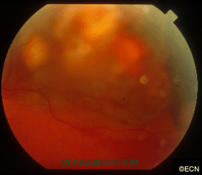
A fundus photograph reveals a marked reduction of subretinal fluid and hemorrhage. Exudates have appeared at the infero-temporal margin, and orange pigment on the inferonasal tumor (as compared to presentation). Note the variably pigmented (mostly amelanotic) subretinal tumor with an associated serous detachment and subretinal hemorrhage.


