Case #18 – Computed axial tomography

Case #18 - Computed axial tomography

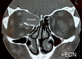
Computed axial tomography demonstrates displacement of the medial rectus muscle (MR), as well as erosion and obliteration of portions of the orbital roof (black arrows). Note that the orbital portion of the mucocele is partially encased in bone. This is characteristic of mucocele. The rectus muscles and optic nerve are labeled.

Patient Stories

"Very well treated by Dr. Finger. He explained everything I needed to know about my issue with detail and attention, putting me at ease and giving me confidence to handle this problem for the rest of my life.”
N.N.

Read More

REQUEST AN APPOINTMENT

Go to Appointment Form

CONNECT AND SHARE