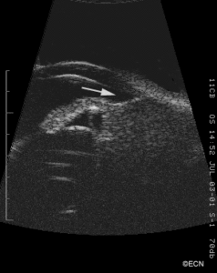
10 and 20 MHz Ultrasonography. High frequency ultrasound (UBM) reveals displacement of the iris root (arrow). Though no sclerostomy is seen, the inner and outer scleral borders are poorly defined. 10 MHz ultrasonography shows an irregularly shaped tumor > 16 mm in diameter.


