By Paul T. Finger, MD
History

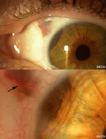
This ciliary body melanoma can cause displacement of the iris root, a sentinel vessel and extrascleral extension. This 38 year-old patient was referred with a large “eye melanoma” with a small plaque of extrascleral extension.
Impression
Anterior Uveal Melanoma with Extrascleral Extension.
10 and 20 MHz Ultrasonography. High frequency ultrasound (UBM) reveals displacement of the iris root (arrow). Though no sclerostomy is seen, the inner and outer scleral borders are poorly defined. 10 MHz ultrasonography shows an irregularly shaped tumor > 16 mm in diameter.
*Note*

This risks and benefits of observation, enucleation and radiation were discussed in detail. Primary enucleation was recommended and performed.
Comment
This case presents several classic findings in anterior uveal melanomas. Please share this case with your physicians in training. It is also important to note that not all ciliary body melanomas will exhibit these findings. Other findings of ciliary body melanomas include sector cataract and irregular astigmatism.
Lastly, there is some controversy about secondary radiation therapy as treatment for presumed residual microscopic orbital melanoma (due to extrascleral extension).
Without any compelling data to suggest its efficacy, and knowing the significant morbidity associated with 50 Gy (typical dose) of external beam radiation therapy to the anophthalmic orbit (dry socket, lash-brow loss, and mucus discharge), no additional treatment was given.
Alternatively, high dose rate intersitial radiation therapy can be used to allow for improved cosmetic results in cases where their is evidence of residual orbital melanoma.


