Chemoreduction of Orbital Tumors

Note the large nodular basal cell carcinoma on the right lower eye lid and cheek prior to cis-platinum chemotherapy.

By Paul T. Finger, MD

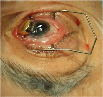
Note the large nodular basal cell carcinoma on the right lower eye lid and cheek in the image below. It has pulled the eye lid out (cicatricial ectropion) and was found to invade the orbit on CT scan. Click on an image below to enlarge and see the full description:

The diagnosis was easily confirmed by taking a small wedge biopsy in the office. Then after a complete discussion of treatment options, this patient opted for intravenous chemotherapy rather than extensive surgery or radiation therapy. In this case, 3 courses of cis-platinum chemotherapy reduced the size of the tumor. Due to renal toxicity, the treatment was discontinued. Chemoreduction did not (in itself) cure this patient, but it did allow for local resection of the reduced sized-tumor. Under frozen section control, tumor-free margins were obtained.

For more information on the investigational technique we offer the following references.

References

  1. Luxenberg MN and Guthrie TH, Jr.. Chemotherapy of eyelid and periorbital tumors. Trans Am Ophthal Soc 1985;83:162-180.

    After 3 courses of treatment the tumor was noted to shrink and his ectropion diminished. Note the improvement in the CT appearance.
    After 3 courses of treatment the tumor was noted to shrink and his ectropion diminished. Note the improvement in the CT appearance.
  2. Luxenberg MN and Guthrie TH, Jr.. Chemotherapy of basal cell and squamous cell carcinoma of the eyelids and periorbital tumors. Ophthalmology 1996;93:504-510.
  3. Morley M, Finger PT, Perlin M, Weiselberg LR, DeBlasio DS. Cis-Platinum Chemotherapy for Ocular Basal Cell Carcinoma. The British Journal of Ophthalmology, 1991, 75, 407-410.

Related Links


Laser Photocoagulation For Radiation Retinopathy

By Paul T. Finger, MD

Dr. Finger uses laser photocoagulation to prevent radiation related retinopathy, maculopathy, and loss of vision. As published in the British Journal of Ophthalmology, Drs. Finger and Kurli found that eyes with posterior choroidal melanomas were more likely to develop sight-threatening radiation retinopathy. In that series, 50 patients were treated with sector scatter laser photocoagulation to clinically evident radiation retinopathy. A second group of patients (considered to be “high risk” to develop radiation retinopathy) were also treated with laser.

In this study, laser photocoagulation improved radiation retinopathy in 29 (64.4%) of the 45 patients treated after the onset of radiation retinopathy (17 with only retinopathy, 10 with a combination of retinopathy and maculopathy, and two with only maculopathy). Of the 16 patients who received laser treatment before clinical evidence of retinopathy, only 1 developed radiation maculopathy and two retinopathy without maculopathy (all three responded to additional laser photocoagulation).
None of the patients in the prophylactic laser group lost more than three lines of vision as a result of maculopathy.

Conclusions

Sector scatter argon laser photocoagulation can be used to induce regression of radiation retinopathy. Though early treatment of radiation retinopathy appears to be more effective, a more long term and prospective randomized study should be performed.

Related Links


18FDG PET/CT SUV: A Noninvasive Biomarker For the Risk of Metastasis from Choroidal Melanoma

By Paul T. Finger, MD

In a research study, Dr. Finger compared the intensity of radioactive glucose uptake [from positron emmission tomography (PET)] to clinical, ultrasound, and pathology features of choroidal melanomas evaluated by FDG positron emission tomography / computed tomography (PET/CT). Ultrasound was used to measure tumor size, evaluate tumor shape and intrinsic vascularity (blood flow). Histopathology and immunohistochemical evaluations of tumor cell-type, necrosis, glycogen-content, vascularity and extrascleral extension were performed.

Selecting out the highest 6 PET/CT standardized uptake values [(SUV) > or = to 4.0] melanomas, patients were (on average) 10 years older. in general, higher SUV tumors had larger basal dimensions, were epithelioid-cell type, were centered anterior to the equator, contained enlarged blood vessels (>150 microns in diameter), and had formed extrascleral extension.

This study suggests that PET/CT imaging offered a physiologic assessment of glucose metabolism within choroidal melanomas. Increased FDG PET/CT SUV ( > or = to 4.0) was positively correlated to known clinical, pathology, and ultrasound features linked to metastatic potential of choroidal melanoma.

Related Links


25 Gauge Incision Anterior Segment Surgery Found Helpful For Both Tumor Biopsy: The Finger Iridectomy Technique

The day after the 1 mm incision "FIT" biopsy, a slit lamp photograph shows the partial thickness biopsy of the tumor.
The day after the 1 mm incision “FIT” biopsy, a slit lamp photograph shows the partial thickness biopsy of the tumor.

Iris, iridociliary, and other anterior segment tumors can be biopsied to help determine if they are a benign or malignant. In the past, a biopsy was either performed by aspiration through a needle [fine needle aspiration biopsy (FNAB)] or by a surgical iridectomy. The advantage of a needle biopsy was the very small “needle” incision that required no stitches or consequences for vision. Unfortunately, it was difficult to do more that scratch the tumor with the needle while aspirating a few cells. Using a sharp needle, the tumor often bleeds, clouding the view and making biopsy more difficult. In the past, the alternative was a surgical iridectomy that required a relatively large corneal incision, removal of a full-thickness piece of iris and sutures to close the corneal wound.

In an effort to make iris and ciliary body tumor biopsy safer and more effective, Dr. Finger invented a technique for minimally invasive iris biopsy (with less surgical trauma). This new technique combines the benefit of 1-mm typically sutureless incision with the ability to retrieve iris tumor biopsy specimens for pathology analysis. He has used this technique to perform tumor biopsy, iridotomy and to remove iris melanoma.

Dr. Finger has also used this technique to treat narrow angle glaucoma. Dr. Finger says, “it would be particularly useful for the treatment of children and mentally challenged adults who are not able to have the laser iridotomy method.” FIT can be used to surgically create a small hole in the iris through a 25 gauge self-sealing incision. “Micro-incision surgery should be safer for this group of patients who might rub their eye after surgery.”

For more information this technique has been recently published in the British Journal of Ophthalmology and Graefes Archive of Clinical and Experimental Ophthalmology.

The Finger iridectomy technique: small incision biopsy of anterior segment tumors.
Finger PT, Latkany P, Kurli M, Iacob C
The British Journal of Ophthalmology 2005;89:946-949

Small incision surgical iridotomy and iridectomy.
Finger PT.
Graefes Arch Clin Exp Ophthalmol. 2005 Aug 2;:1-2

The Finger Iridectomy Technique for Glaucoma
Finger PT
British Journal of Ophthalmology 2007;91:1089-1090.

Related Links


Amniotic Membrane Is Used To Protect The Cornea During Plaque Radiation Therapy

Amniotic Membrane Graft on the Cornea (Arrow) affixed beneath a Custom Gold Eye Plaque

By Paul T. Finger, MD

Patients with melanomas of the iris and ciliary body are more and more commonly treated with plaque radiation therapy. This is because I found radiation to be less invasive and therefore safer than intraocular surgery. Plus, larger areas can be treated with radiation than can be safely removed. This has allowed for excellent local control (tumor destruction) and vision retention. Though many irradiated patients develop radiation cataracts, these can be removed (the same way as other cataracts) restoring excellent vision. At The New York Eye Cancer Center, almost no patients with anterior intraocular melanomas have developed sight-limiting radiation maculopathy or optic neuropathy.

However, in the past, there have been problems associated with placing the radioactive plaque onto the cornea. First, there were concerns about irradiating the cornea. Second, many patients found having a metal device sewn to their cornea for 4-7 days painful.

Dr. Finger has found the cornea tolerated the amount of radiation required to treat iris and ciliary body melanomas. It remained clear for the vast majority of patients. However, having a radiation plaque sewn to the cornea was an uncomfortable procedure.

In an effort to solve this problem, Dr. Finger discovered that a thin, transplanted amniotic membrane tissue could be placed between the gold radiation eye plaque and the cornea during treatment. He simply slides this tissue between the plaque and the cornea during implantation. It is removed 7 days later when the plaque is removed. This technique has made a huge difference – it improved patient comfort and does not affect the radiation treatment.

This note is linked to a You-Tube video demonstrating this new technique. It shows implantation of a plaque onto the cornea and eye wall.

Related Links


Whole Body FDG – PET / CT: Imaging Cancer

By Paul T. Finger, MD

Abdominal Section: Note that the fusion of the black and white forms seen on CT with the color (metabolic activity seen on PET) reveals liver metastasis.

Whole body PET/CT technology combines positron emission tomography (PET) with computed radiographic imaging (CT) to put FUNCTION and FORM on the same diagnostic page (PET/CT).

Spiral computed tomography CT is used to generate anatomic images of the entire body. When suspicious areas or tumors are found, CT allows your doctor to see their size, shape and internal radiographic density. Though computed radiographic tomography (and magnetic resonance imaging – MRI) are excellent methods to determine if an abnormality exists, its shape and location, it cannot reveal if the abnormality (e.g. tumor, lesion) is metabolically active.

In contrast, Positron Emission Tomography (PET) is used to determine if tissues or tumors are metabolically active. This is important, because metabolically active tumors are more likely to be malignant. In the case of imaging of malignant melanoma, PET imaging can differentiate between benign cysts and metastatic tumors in the liver (and other sites). PET requires a small injection of radioactive material (e.g. FDG – radioactive glucose) that is preferentially absorbed by malignant tumors. It is important to note that any glucose absorbing process (inflammations, infections, working muscles and excretory systems) will also concentrate glucose and appear as a “hot-spot” on PET. However, PET does not give you shape or location. That is why and additional CT is needed. It is the addition of the anatomic information provided by CT that allows the physician to differentiate between benign and malignant tumors. The PET/CT computers unify the PET information over the CT information, placing form and function on the same diagnostic page.

Dr. Finger has shown the though the liver is the most common initial site of metastatic choroidal melanoma. It can occur in the bones (in 50% of cases). Clearly, a whole-body PET/CT scan from the top of the head to the bottom of the feet will include all the bones in the body. It is important to realized that not all metastases will “light-up” with PET and that sometimes a dedicated CT or MRI will offer better anatomic imaging. So don’t be surprised if there is suspicious area seen on PET/CT, your doctor may request an additional radiographic examination.

We expect more melanoma specific radioactive materials (other than FDG – radioactive glucose) to be used for PET/CT. This will improve the specificity and sensitivity of this test. To see the research work being conducted thus far, visit the links below.

Related Links


3-Dimensional Ultrasonography

Oblique sections demonstrate high reflectivity of the plaque's posterior surface.
Oblique sections demonstrate high reflectivity of the plaque’s posterior surface.

By Paul T. Finger, MD

3D ultrasound has been used to image radioactive eye-plaques while they are sewn beneath their intraocular tumors. This technique was used to make sure radioactive eye plaques are properly positioned beneath its intraocular tumor.

3D ultrasound is particularly helpful when examining eyes with tumors, retinal detachment and calcifications (e.g. retinoblastoma). It is a relatively inexpensive way to measure the diameter of the orbital portion of the optic nerve.

References

Finger PT, Romero JM, Rosen RB, Iezzi R, Emery R, Berson A. Three-dimensional ultrasonography of choroidal melanoma.Localization of ophthalmic plaques. The Archives Ophthalmology 116:305-12, 1998.

Related Links


Patient Stories

"Very well treated by Dr. Finger. He explained everything I needed to know about my issue with detail and attention, putting me at ease and giving me confidence to handle this problem for the rest of my life.”
N.N.

Read More

REQUEST AN APPOINTMENT

Go to Appointment Form

CONNECT AND SHARE