New Research Points to Role of Immune System in Fighting Uveal Melanoma

A study published in August in JAMA Ophthalmology probes the importance of inflammation associated with immune system response in uveal melanoma.

Uveal melanoma is an eye cancer that involves the iris, the muscle surrounding the lens, or the choroid – a vascular layer with connective tissue between the retina and the sclera. It is generally well-controlled by radiation therapy or surgery, but often develops aggressive metastases. Once the cancer spreads to other parts of the body, there are very few cures.
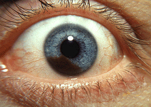
iris_melanoma

Researchers have found that certain genetic alterations are associated with metastases, but gain in a specific chromosome – 6P – is associated with a more favorable outcome.

Ulrich Pfeffer, PhD with the Laboratory of Molecular Pathology Istituto di Ricovero e Cura a Carattere Scientifico Azienda Ospedaliera Universitaria San Martino in Genoa, Italy, said many have guessed that the immune system, or more generally, inflammation might protect uveal melanomas with this alteration from progression to metastasis.

Seeking to better understand the process, researchers looked at a gene on the 6P chromosome, the putative immunomodulatory BTNL2. An immunomodulator is a chemical agent that modifies the immune response or the functioning of the immune system. Pfeffer described the findings:

“We found highly variable expression of this gene in uveal melanoma samples where it is expressed by tumor cells and by infiltrating immune cells. The type of infiltrate is strongly associated with the risk to develop metastases.”

While the exact operation of BTNL2 and its interaction with the immune system remains unclear, the research does indicate a relationship between immune response and metastasis in uveal melanoma. Pfeffer said this is key:

“Inflammation is an important feature of uveal melanoma and BTNL2 is likely to be an important determinant of the local immunological environment.”

He recommends further research in the hopes effective immunotherapies can be developed:

“BTNL2 is a member of a family of genes that share many features with other immunomodulatory genes. Some of these genes are targets for highly successful therapies. However, many uveal melanoma patients do not respond to these therapies. Future research should address BTNL2 as a specific modulator of the immunological niche of the eye.”

Inflammation is often associated with intraocular melanoma. It can be seen both prior to or after radiation treatment. Signs of inflammation suggest that the patient’s own immune system is trying to reject or destroy the primary cancer.

Another study we reported on last spring found a subset of T-cells in uveal melanoma that had robust anti-tumor reactivity. With further research, it may be possible to build on this knowledge to discover therapies that will boost the immune system’s response and/or effectiveness to successfully treat uveal melanoma.

The studies also underscore the importance of boosting the immune system generally when fighting eye cancer. There are a number of simple steps patients can take to strengthen the immune system, including:

  • Eating a healthy, well-balanced diet.
  • Getting plenty of sleep.
  • Moderate exercise.
  • Avoiding excessive stress.

Harvard Health Publications has more detailed information on steps to boost the body’s immune system.

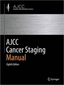
8th Edition AJCC Cancer Staging Completed

The American Joint Committee on Cancer (AJCC) recently celebrated the completion of the 8th edition of its Cancer Staging Manual.

AJCC Cancer Staging Manual, 8th EditionThe AJCC Cancer Staging Manual defines an international language for oncology. Created with and adopted by the Union for International Cancer Control (UICC), the AJCC-UICC TNM Cancer Classifications enables communication between eye cancer care providers all over the world. The manual brings together all currently available information on staging of cancer, and incorporates newly acquired knowledge on cancer etiology and pathology.
The AJCC Cancer Staging Manual:

  • Enables communication within the local health care team.
  • Makes patient education more effective.
  • Improves clinical and patient-related decision making.
  • Allows for quality measurements.
  • Improves clinical research.

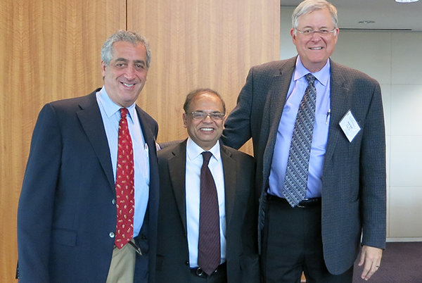
On September 9, a meeting was held at the American College of Surgeons headquarters in Chicago, Illinois to celebrate the completion and publication of the Manual. It was the culmination of a 5-year effort to create the best possible evidence-based cancer classification system.

Dr. Finger at AJCC Meeting
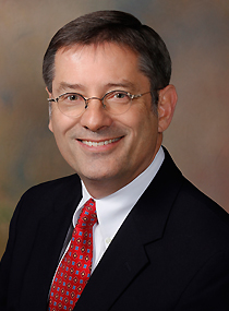
Paul T. Finger MD (Chairman of The Eye Cancer Foundation), Mahul B. Amin MD (Editor-in-Chief of the AJCC) and David R. Byrd MD (Chairman of the AJCC)

Paul T. Finger, MD attended the meeting, representing the 54 Ophthalmic Oncology Task Force (OOTF) members who volunteered countless hours toward making the best possible, consensus-based eye cancer staging system. As Chair of the OOTF, Dr. Finger was primarily responsible to the AJCC for eight chapters submitted for publication.

“This is a unique text that offers the best possible consensus of medical fact and opinion, and most importantly it has been generated by a broad representation of our subspecialty,” Dr. Finger said. “We covered all aspects of the diagnosis and treatment of eye cancer. Everyone who cares for patients with eye cancer should read this book.”

The AJCC is comprised of 20 members including the American Cancer Society, the National Cancer Institute, the American Society of Clinical Oncology and other organizations focused on the diagnosis and treatment of cancer.

The 8th edition AJCC staging manual is available for pre-order on Amazon.

Members of the Ophthalmic Sites Expert Panel

Daniel M. Albert, MD, MS

Anush G. Amiryan, MD

Claudia Auw-Hädrich, MD

Diane Baker, CTR

Raymond Barnhill, MD, MSc

José M. Caminal, MD, PhD

William L. Carroll, MD

Nathalie Cassoux, MD, PhD

Jaume Catalá-Mora, MD

Guillermo Chantada, MD

Patricia Chévez-Barrios, MD

R. Max Conway, MD, PhD, FRANZCO

Sarah E. Coupland, MBBS, PhD – Vice Chair

Bertil E. Damato, MD, PhD

Hakan Demirci, MD

Laurence G. Desjardins, MD

François Doz, MD, MSc

Jonathan J. Dutton, MD, PhD, FACS

Bita Esmaeli, MD, FACS

Paul T. Finger, MD, FACS – Chair

Brenda L. Gallie, MD, FACSC

Gerardo F. Graue, MD

Hans E. Grossniklaus, MD – CAP

Steffen Heegaard, MD

Leonard M. Holbach, MD

Santosh G. Honavar, MD, FACS

Martine J. Jager, MD, PhD

Tero Kivelä, MD, FEBO

Emma Kujala, MD

Livia Lumbroso-Le Rouic, MD

Ashwin C. Mallipatna, MBBS, MS, DNB

Giulio M. Modorati, MD

Francis L. Munier, MD

Timothy G. Murray, MD, MBA, FACS

Anna C. Pavlick, MS, DO

Jacob Pe’er, MD

David E. Pelayes, MD, PhD

Gaelle Pierron

Victor G. Prieto, MD, PhD

Manuel Jorge Rodrigues, MD

Svetlana Saakyan, MD

Wolfgang A.G. Sauerwein, MD, PhD

Ekaterina Semenova, MD

Stefan Seregard, MD

Carol Shields, MD

E. Rand Simpson, MD, FRCS(C)

Arun D. Singh, MD

Shigenobu Suzuki, MD, PhD

Mary Kay Washington, MD, PhD

Valerie A. White, MD, MHSc, FRCPC

Michelle Williams, MD

Matthew W. Wilson, MD, FACS

Christian W. Wittekind, MD – UICC

Vivian Yin, MD, MPH, FRCSC


ECF’s Second Working Day Now Part of Official ISOO Biennial Conference

The Eye Cancer Foundation is gearing up for the Second Working Day, which is now part of the official ISOO Biennial Conference. It will commence on Friday, March 24, 2017 at the International Convention Centre in Sydney, Australia.

If you’re an eye cancer specialist attending the conference, be sure to mark your calendars and arrive by Thursday night!

The first Working Day in Paris was a resounding success and generated a lot of positive energy that carried over into the official ISOO Conference. It featured 6 sections:

  • Eye cancer registries (data mining to provide medical evidence)
  • Retinoblastoma fellowships (outreach to the un-served)
  • Surgical standards (outreach)
  • Radiation side effects as well as patient and doctor reported outcomes.
  • Patient reported outcomes
  • Doctor reported outcomes

Due to time constraints in Sydney, work will be continued on only 4 sections. These ongoing projects strive to create consensus and maximum participation from our members of the ISOO.

2017 Working Day will feature five separate committees on the following topics:

  • International Medical Evidence (WD-ME), Lead by Bita Esmaeli MD. ISOO members will be made aware of all the international multi-center, data-driven projects that have been completed, those in progress, and others just starting. They will discuss international medical evidence committee standards for publication, participation in ongoing registries, new projects, and funding.
  • Retinoblastoma Fellowships (WD-RBF), Lead by Ashwin Mallipatna MD. ISOO members will discuss the RBF Committee’s work on what constitutes basic educational standards for our retinoblastoma fellows and strategize how to support their work as they return to their underserved countries.
  • Quality Assurance (WD-QA), Lead by Tero Kivela MD. Ten centers will voluntarily share their outcomes. ISOO members will discuss these efforts and discuss how to move forward.
  • Surgical Standards (WD-SS), Lead by Santosh Honavar MD. ISOO Members will strategize on how Dr. Honavar’s committee text will be published online with free access. Thus, our community will provide consensus surgical guidance to all the general ophthalmologists currently treating eye cancer patients.
  • Consensus Guidelines (WD-CG), Lead by Paul T Finger MD. One designated representative from each eye cancer center will receive a free copy of the newly published 8th edition, AJCC Staging Handbook. All 70 contributors will be acknowledged and ongoing AJCC-UICC initiatives and other consensus guideline initiatives will be introduced for construction.

Smartphone Technology Opening Doors for Vision Impaired Patients

The advent of smartphones and tablets has opened up new doors for patients with vision loss. In the past, they had to deal with heavy, expensive hardware in order to use assistive technology. Today, patients with vision loss can access inexpensive apps on lightweight devices that help them gain independence and better navigate the world around them.

Mobile apps for vision aid

Some standard features on an iPhone and iPad are invaluable for low-vision patients. Siri’s voice activated technology allows visually impaired people to surf the web and access many of the phone’s features. The camera also provides an aid, allowing users to take photos and magnify them in order to see details they otherwise couldn’t make out. You can review all of iOS’ accessibility features here.

Beyond these basic features, there are also a number of low-cost apps made specifically for low-vision users.

Even simple apps for general use can prove helpful to those with vision impairment. For instance, a variety of apps display a large digital clock on the phone that is much easier for a low-vision user to see. A headphone app called Awareness! allows the user to hear surrounding sounds through their headphones even while listening to music. This is invaluable for people who depend on their ears to navigate.

Beyond these basic applications, there are a number of apps made specifically for visually impaired users.

Blindsquare for the iPhone helps with outdoor navigation. It uses a dedicated speech synthesizer and interprets real-time GPS data from FourSquare and the Open Street Map database to describe the surrounding area, announce intersections, and identify points of interest.

LookTel Recognizer is another app that can help with day-to-day activities. It identifies items such as food packages, DVDs, and credit cards using the phone’s camera. The app compares the item with a user-generated database of photos stored in the phone itself.

CamFind was developed to assist with comparison shopping, but sight-impaired users have repurposed it to help identify various objects. You simply take a photo, and the app compares it to known Internet images and announces what the object is.

An iOS app called TapTapSee serves a similar function. The user double-taps the screen and the phone takes a photo. A synthesized voice gives an ID of the object.

KNFB Reader allows the user to “read” signs, menus, or any written document simply by taking a photo. The app uses text-to-speech technology to read it out loud.

List Recorder allows users to record and organize lists using either audio or text. It also integrates with voiceover or braille displays.

AccessNote and Notablilty are both good note-taking apps that integrate text and voice. Notability also allows the user to manipulate photos. You can zoom in for better viewing and add audio notations.

This is just a sampling of the low-cost assistive technology available for the vision impaired. There are several websites available with more information.

  • AppleVis is a website published by sight-impaired users that provides information about assistive apps. The site includes a directory of smartphone apps and games with user reviews.
  • The Eyes-Free Project has developed several free low-vision apps for Android smartphones and tablets. You can find them in the Google Play store.

New Study Shows Effectiveness of Minimally Invasive Biopsy Technique Pioneered by Dr. Finger

Dr. Paul Finger and Dr. Tatyana Milman’s recently published study in the European Journal of Ophthalmology highlights the effectiveness of an innovative minimally invasive biopsy technique.

Biopsy-proven iris melanoma carries a 10.7% risk of metastasis, making a safe method of iris tumor biopsy crucial. Currently, physicians often use fine-needle aspiration or wide-incision iridectomy. However, both techniques have drawbacks.

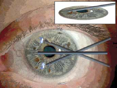
With fine-needle aspiration, piercing or partially embedding a sharp-edged needle tip into a 0.8-mm thick iris risks cutting the lens, iris blood vessels, and ciliary body. Surgical iridectomy yields the best tissue samples, but involves creating a relatively large surgical wound that must be sutured. This risks astigmatism, an irregular pupil, secondary glare and the need for post-op rehabilitation.

As the new study shows, using a microincision aspiration cutter is a third, minimally invasive option that yields excellent biopsy results while minimizing some of the risks associated with the other two methods.

microincision aspiration cutter

Microincision aspiration cutter-assisted iris biopsy is a variation of the Finger iridectomy technique developed by Dr. Finger. It uses a single, small corneal incision, which is safer than the larger incision required by wide-incision iridectomy. Dr. Finger explained the significance of this advancement in biopsy techniques for patients:

“Conducting a biopsy through a tiny incision like this means you don’t have to have stitches. Think of it like the developments in back or abdomen surgery over the past few decades. Surgeons used to have to really open the patient up, which often meant more pain and a longer post-op recovery. Then they developed microincisions to conduct surgery. This is the ophthalmic version of those surgical enhancements – a surgery upgrade, if you will.”

In this study, seven patients suspected to have diffuse iris melanoma underwent microincision aspiration cutter-assisted iris biopsy. Five of the patients were ultimately diagnosed with melanoma. None of the patients lost visual acuity, developed biopsy-related glaucoma, nor experienced hypema (bleeding in the eye). In fact, 75% of the patients showed reduced intraocular pressure after the procedure.

Microincision aspiration cutter-assisted iris biopsy can provide doctors with a safer alternative to the other two commonly used methods. It allows effective biopsies with a small incision, rapid rehabilitation and potentially fewer complications.


UK Patient’s Story Shows Importance of Routine Eye Exams for Cancer Detection

Ralph McMurray of Suffolk, England, was 66 years old when he had his first ever eye examination.

It revealed he had eye cancer.

West Suffolk Hospital

According to a local news report, the examining optometrist found a detached retina and referred McMurray to the West Suffolk Hospital for further tests, which revealed a large tumor in his eye – an ocular melanoma. Physicians at St. Bartholomew’s Hospital in London removed McMurray’s eye just weeks later due to the size of the tumor.

“I am so glad I went for that eye test as the cancer would have only got worse and I may not even be here now,” said McMurray. “I am now determined to do as much as I can to raise awareness of the importance of having regular eye tests… In cases such as mine early diagnosis is crucial and the tumor would have been spotted much sooner had I been having regular eye examinations.”

McMurray’s experience underscores the importance of routine eye exams and the importance of all eye care professionals learning Dr. Fingers’ MOST mnemonic (Melanoma = Orange pigment, Subretinal Fluid, Thickness of 2 or more millimeters) to effectively smaller intraocular melanomas.

The earlier a tumor is detected, the more likely vision- and life-saving treatments can be employed.

McMurray was fortunate the cancer was found, even though it had already advanced. Prompt treatment after diagnosis likely saved his life.

For UK nationals considering a second opinion or treatment in the United States, The New York Eye Cancer Center offers special programs to assist international patients. Click here to learn more.


ICO/ECF Join Forces to Treat Childhood Eye Cancer in Underserved Countries

Proper Training Expected to Substantially Reduce Mortality Rates 

The Eye Cancer Foundation is pleased to announce a partnership with the International Council of Ophthalmology (ICO) to promote and conduct eye cancer fellowship education for physicians from unserved countries.

The Eye Cancer Foundation and International Council of Ophthalmology

The ICO represents and serves professional associations of ophthalmologists from around the world. The organization’s mission is to “work with ophthalmologic societies and others to enhance ophthalmic education and improve access to the highest quality eye care in order to preserve and restore vision for the people of the world.”

The ICO Fellowship program leadership team
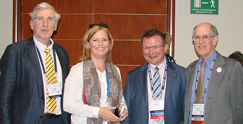
Left to right: Peter Gabel, MD (former ICO Fellowships Director), Cordula Gabel-Obermaier, MD (ICO Fellowships Executive), Berthold Seitz, MD (ICO Director for Fellowships), and Bill Felch (ICO CEO).

This new partnership will expand and enhance The Eye Cancer Foundation’s existing fellowship programs. It will focus specifically on addressing the need to reduce world-wide mortality related to retinoblastoma by ensuring that more patients have ready access to a trained eye cancer specialist in their region.

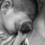
Untreated childhood retinoblastoma

Retinoblastoma is the most common eye cancer in children. It typically develops by the age of five. Retinoblastoma affects approximately 8,200 children each year. The incidence rate is somewhat higher in developing countries, where most of the children succumb to metastatic retinoblastoma. In contrast, there exists a better than 96% survival rate in developed countries like the USA.

Early diagnosis and prompt treatment will save lives. In countries without eye cancer specialists, children with retinoblastoma typically go undiagnosed and untreated until it is too late. Local general ophthalmologists are not familiar with modern eye cancer medicine. Training doctors to diagnose and treat retinoblastoma in these areas will save lives. The Eye Cancer Foundation and the International Council of Ophthalmology has set a goal of saving 1,000 children by 2020.

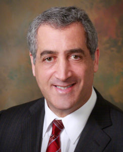
Paul T. Finger, MD
Dr. Paul T. Finger, Chairman, The Eye Cancer Foundation

The Eye Cancer Foundation currently funds 25 half-year retinoblastoma fellowships in ophthalmic oncology for candidates from unserved or underserved countries. The new partnership with the ICO will help expand the scope and reach of this fellowship program.

“The Eye Cancer Foundation is looking forward to a long and productive relationship with the International Council of Ophthalmology,” says our Chairman Dr. Paul Finger


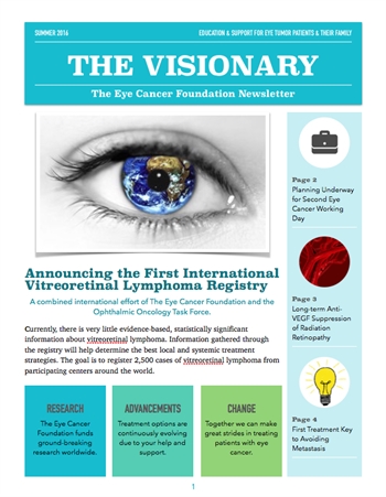
The Visionary Newsletter – Summer 2016 Edition

The following was originally published by The Eye Cancer Foundation.

The Visionary Newsletter - Summer 2016 EditionThe Summer 2016 edition of The Visionary is now available! The Eye Cancer Foundation publishes The Visionary free-of-charge to keep you informed about the latest news, research, and global efforts focused on improving eye cancer treatment, diagnosis, and cure.

In this edition, you will find articles about:

  • The First International Vitreoretinal Lymphoma Registry
  • The Second Eye Cancer Working Day in 2017
  • A Massive International Study Yielding Key Insights into Preventing Metastasis
  • And More

The Eye Cancer Foundation can send The Visionary directly to your inbox. If you’d like to receive a personal email notification when new editions of The Visionary are released, please follow this link to the eyecancercure.com website. Your information will not be shared with third parties.


New ECF-Sponsored Ophthalmic Pathology Fellowship at Emory Eye Center with Hans Grossniklaus, MD, MBA

dr_grossniklaus
Hans Grossniklaus, MD, MBA, founding director of the Ocular Oncology and Pathology Services at Emory Eye Center in Atlanta, Georgia

We are fortunate to have partnered with Hans Grossniklaus, MD, MBA who is founding director of the Ocular Oncology and Pathology Services at Emory Eye Center. Through this partnership, The Eye Cancer Foundation (ECF) is continuing its work supporting fellowship education for doctors from unserved countries.

Dr. Hans E. Grossniklaus will host one ECF fellow per year from an unserved country or area. The fellowship will focus on ophthalmic pathology, and doctors completing the program will return to practice in their home country. These new eye pathologists will be trained to provide crucial eye pathology services for our emerging retinoblastoma fellows.

Ophthalmic pathology is the subspecialty of surgical pathology and ophthalmology that primarily deals with the diagnosis and characterization of cancer and non-cancerous diseases of the eye, eyelids and orbit. Ophthalmic pathologists are vital in providing practicing ophthalmologists with information about causes, pathogenesis, and the prognosis of ocular diseases.

The new ECF fellowship will provide a $30,000 grant for one year of travel and expenses. Candidates must have completed a residency in ophthalmology. For consideration, they will need to submit a letter of recommendation from their local chairman or hospital director. Included in this letter must be a clear statement that the prospective fellow will be supported to perform ophthalmic pathology upon return to their home country. Failure to return home would make the fellow liable to repay the grant money.

The Emory Eye Center is the largest, most comprehensive clinical eye care facility in the southeast United States. Dr. Grossniklaus is widely recognized as a national leader in eye disease treatment and education, as well as a pioneer in eye disease research. Learn more here.

This ECF-sponsored ophthalmic oncology fellowship provides doctors training they would be unable to get in their home country. Their presence will reduce mortality and save vision in countries that currently lack the resources to effectively diagnose and treat many eye diseases, including eye cancer.

If you are a candidate in an unserved country interested in the fellowship, or would like to recommend a candidate, please contact The Eye Cancer Foundation at (212) 832-8170 or click here to email us.


ECF Fellow Publishes Uveal Melanoma Study in Middle East

Dr. Ahmad Shokri Zewar from Yemen was the first author of a recent study to evaluate the features and outcomes relating to the management of uveal melanoma in King Hussein Cancer Center in Jordan. Zewar conducted the study with Dr. Yacoub A.Yousef, a collaboration that was possible thanks to The Eye Cancer Foundation’s Fellowship program that supports training of eye cancer specialists from unserved countries.

Dr. Ahmad Shokri Zewar & Dr. Yacoub A.Yousef

While the paper focused specifically on King Hussein Cancer Center, the aim was to complete a study that could be generalized to other referral tertiary cancer centers in the Middle East.

The study focused on 46 patients with uveal melanoma between 2006 and 2014. There were actually 76 patients diagnosed in the hospital over that time period, but 30 were excluded from the study due to lack of data or because they didn’t receive follow-up. This is a relatively low number, and researchers concluded it provides the impression that uveal melanoma occurs with a low frequency in the Middle East as compared to Western countries.

Eighteen eyes (39% of the patients) were treated by primary enucleation, and 28 eyes (61%) were treated by I-125 radioactive plaque therapy. Two of the eyes treated by plaque therapy were subsequently enucleated. Many of the patients were not diagnosed until the disease had significantly advanced.

The authors write:

“In our review, a significant percentage (65%) of affected eyes had a visual acuity of less than 0.1, which is worse than the visual acuity for of patients studied in the COMS study where only 33% had visual acuity of less than 0.1 (9). This finding might be explained by the delay in presentation after the onset of ocular complaints in developing poor countries where health care could be unachievable because of the high cost or of far distance to travel.”

Late diagnoses lead to a higher number of enucleations and more metastatic melanoma:

“A significant number of Arabic patients, unfortunately, present to ocular oncology clinics at a time where the tumor reaches a large size or is associated with complications that make it non-amenable for brachytherapy and end up with enucleation. Therefore, awareness must be increased and early detection improved with prompt referral by the general ophthalmologist to save more eyes and to enhance survival of affected patients.”

Remember, to effectively diagnose Choroidal Melanoma, ophthalmologists just need to remember “MOST.”

The paper could serve as a springboard for more aggressive efforts to raise awareness of uveal melanoma and its symptoms. This could lead to early diagnoses and a higher cure rate without the loss of an eye.

The goal of the ECF Fellowship Program is to help train candidates from underserved countries so they can return and establish programs for the local populace. The fellowship provides training, a stipend, and travel expenses. In return, doctors agree to return to their home country and treat eye cancer patients.

Drs. Ahmad Shokri Zewar and Yacoub A.Yousef’s work demonstrates the value of investing in doctors in underserved areas and attest to the success of the Eye Cancer Foundation fellowships.


Patient Stories

"Very well treated by Dr. Finger. He explained everything I needed to know about my issue with detail and attention, putting me at ease and giving me confidence to handle this problem for the rest of my life.”
N.N.

Learn More About Our Patient Care

REQUEST AN APPOINTMENT

Go to Appointment Form

CONNECT AND SHARE