
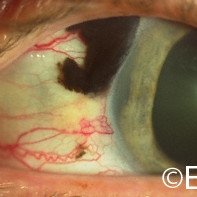
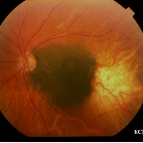
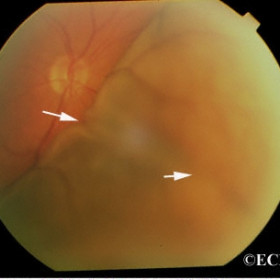
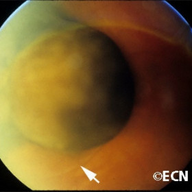
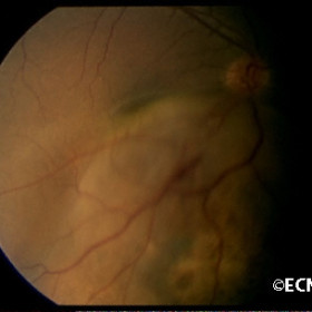
Ciliary body melanoma
Sentinel (Def- a thing that indicates the presence of disease) vessels overlying the melanoma (arrow).
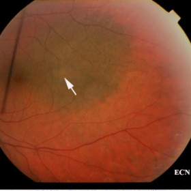
Choroidal Nevus
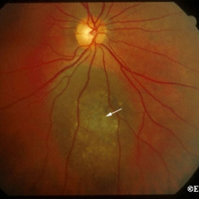
This suspicious choroidal nevus demonstrates thinning of the retinal pigment epithelium and subretinal fluid.
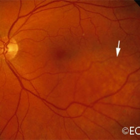
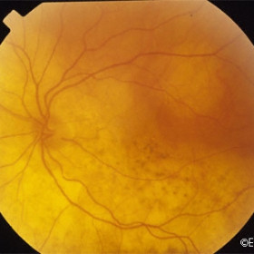
Choroidal Nevus
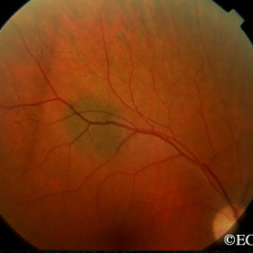
This relatively benign appearing nevus has no evidence of orange pigment, subretinal fluid or appreciable thickness.
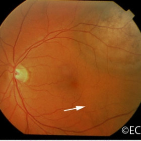
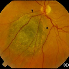
Choroidal Nevus
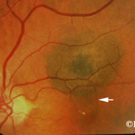
The suspicious choroidal nevus and its secondary retinal detachment are both located in the macula.
Choroidal Nevus
This suspicious choroidal nevus presented with overlying CNV, was treated with observation for 3 years with no growth or vision loss (20:40)
Choroidal nevus, Blue-domed
Thinning of Bruch`s Membrane and the retinal pigment epithelium (arrow).
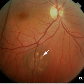
Choroidal nevus
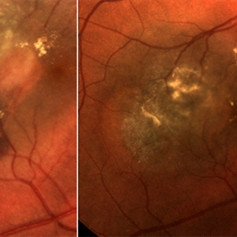
Choroidal nevus with drusen (arrow)
Choroidal osteoma
Choroidal osteoma- Peripapillary with hemorrhage at the macular margin suggesting associated subretinal neovascularization
Choroidal osteoma
Choroidal osteoma - Fluorescein angiography demonstrates subretinal neovascularization at the macular margin.
Choroidal osteoma
Choroidal osteoma with scalloped edges, pigment on surface and yellow coloration.
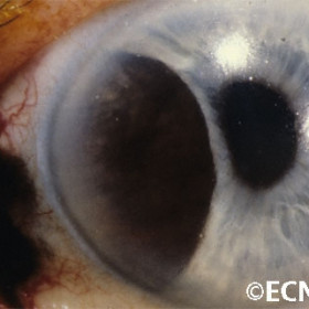
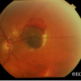
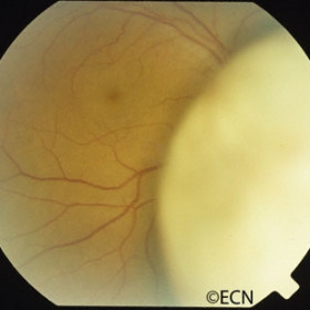
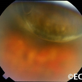
Ciliary body melanoma
Ciliary body melanoma behind the iris as seen through a dilated pupil
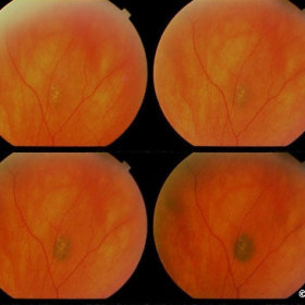
Choroidal nevus
These photographs (taken during the same session) demonstrate how lighting can affect the apparent size of a choroidal nevus.
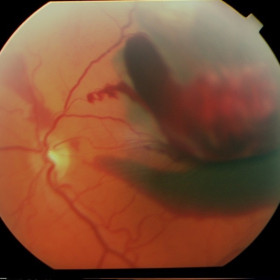
Ciliary Body Melanoma
Ring melanoma, with 360 degrees of sentinel vessels, ectropion, and superior extrascleral extension.
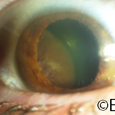
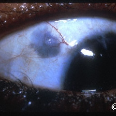
Ciliary body melanoma
Ciliary body melanoma with iris displacement and extrascleral extension.
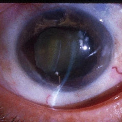
Ciliary body melanoma with iris neovascularization
Ciliary body melanoma with iris neovascularization, displacement of the lens and secondary glaucoma.
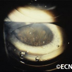
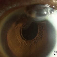
Ciliary body melanoma
Ciliary body melanoma as seen on gonioscopy- Note the tumor growing through the ciliary body band.
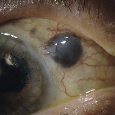
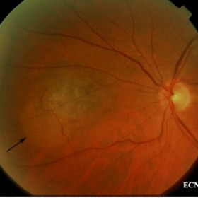
Ciliary body melanoma extension
Note the conjunctival blood vessels draped over the tumor.
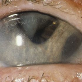
Ciliary body melanoma with extrascleral tumor extension
Ciliary body melanoma with extrascleral tumor extension, disinsertion of the iris root and flattening of the pupillary margin.
Interferon retinopathy
Interferon retinopathy seen in a patient treated for metastatic choroidal melanoma.
Scleromalacia...
Scleromalacia can be confused with scleral extension of uveal melanoma
Uveal prolapse
Note the updrawn pupil in this case of %22pseudomelanomatous%22 uveal prolapse. High frequency ultrasound was diagnostic.
Choroidal Melanoma
The melanoma related exudative retinal detachment is noted to form a demarcation line (arrow).
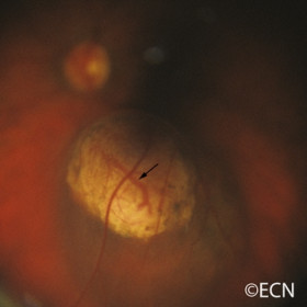
Choroidal hemangioma, circumscribed
Choroidal hemangioma, circumscribed (arrow) in a juxtafoveal position.
Choroidal Hemorrhage
Note the fresh hemorrhage and exudates at this pigmented lesions periphery.
Choroidal Hemorrhage
Choroidal melanoma
Choroidal melanoma - Juxtapapillary, now regressed with chorioretinal atrophy now 2 years after palladium-103 plaque radiation therapy.
Choroidal melanoma
Choroidal melanoma - T1:AJCC small with subretinal fluid (arrow), orange pigment and thickness of 2 mm.
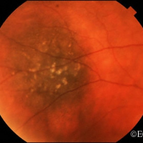
Choroidal melanoma
Choroidal melanoma - Orange Pigment (arrows)
Choroidal melanoma
Choroidal melanoma, broken through Bruch`s membrane as to expose intrinsic vascularity (arrow). Orange pigment and a localized retinal detachment are noted.
Choroidal melanoma
T3:COMS large overhanging the optic nerve
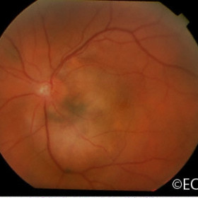
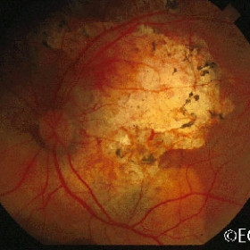
Choroidal Melanoma
Juxtapapillary variably pigmented choroidal melanoma.
Choroidal Effusion
Choroidal Effusion- Note the retinal folds (arrows) suggestive of choroidal effusion.
Choroidal Melanoma
This amelanotic (non-pigmented) choroidal melanoma was uncovered after surgical clearing of a vitreous hemorrhage (vitrectomy).
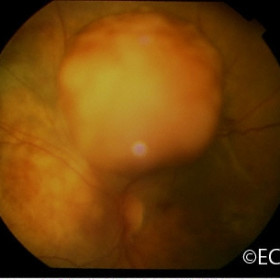
Choroidal melanoma
Collar button shaped also called "mushroom shaped" with a small associated retinal detachment (arrow).
Choroidal melanoma
Choroidal melanoma with orange pigment (arrows).
Choroidal melanoma
Anterior location- Note that the pars plana raised over the tumor`s surface.
Choroidal metastasis
Thyroid cancer metastatic to the juxtapapillary choroid of the eye.
Choroidal metastasis
Choroidal metastasis- Wilm`s tumor origin
Choroidal metastasis
A clinical photograph of a choroidal metastasis from a primary breast cancer showing early response to external beam radiation therapy.
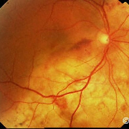
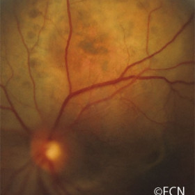
Choroidal nevus
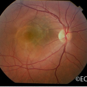
Large, juxtapapillary with a small amount of orange pigment on its surface (arrow).
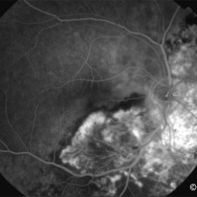
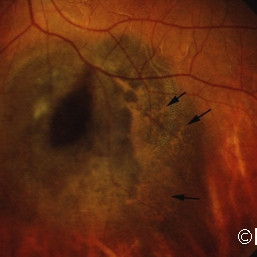
Choroidal Nevus
Note the small exudative retinal detachment along the inferior margin of this suspicious choroidal nevus.


