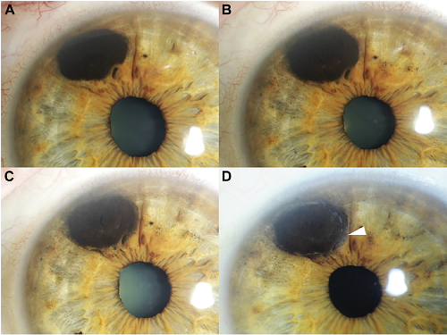
Iris Biopsy 101: The Finger Iridectomy Technique


In order to examine the existence and extent of an iris tumor, a biopsy may be required — but what is a biopsy, exactly? For those who may be unfamiliar with the term, a biopsy is a sampling of tissue taken from a certain area of the body in order for doctors to closely evaluate a patient’s pathology. Every biopsy is different. However, they can sometimes be invasive and cause discomfort. For a patient undergoing a biopsy in an area as delicate as the eye, this may feel intimidating.

This important factor has contributed to Dr. Finger’s development of the Finger Iridectomy Technique (F.I.T), a minimally invasive but very effective method of biopsy. This technique allows for a large amount of tissues and cells to be biopsied, optimizing sample size while minimizing discomfort for patients.

In this video, Dr. Krystine Olszewski from the New York Eye Cancer Center provides an approachable checklist on what to do before and after an iris biopsy. She discusses the required medication to be taken, as well as what is to be expected in the follow up exam.

Iris Biopsy; Finger Iridectomy Technique from Paul T Finger on Vimeo.

To stay tuned for upcoming videos as well as be informed on all the latest updates in eye cancer research, keep Eyecancer.com, the website for the New York Eye Cancer Center, in your bookmarks!


Get to Know: Dr. Paul T. Finger

The New York Eye Cancer Center is dedicated to combining both science and technology with a human touch, offering a holistic approach to the diagnosis and treatment of eye cancer. The Founding Director, Dr. Paul T. Finger, aims to extend his streamlined medical approach to others with the focus of not only helping doctors work together, but also providing easy-to-understand information. This is very important for patients who feel overwhelmed by the scope of medical terminology and technology. Every day, swift strides are being made towards a brighter future for eye cancer patients all around the world.

Here at The NYECC, a series of videos have been made and published, ranging from lectures for training specialists, to videos for patients on what to expect throughout the stages of their care.

In this video, Dr. Finger timelines his 30+ year medical career. He discusses his pioneering of the Palladium-103 plaque procedure for intraocular tumors, imaging methods for many eye cancers and anti-VEGF therapy now used world-wide to suppress radiation complications, plus more.


Meet Dr. Paul T. Finger, Director of The New York Eye Cancer Center from Paul T Finger on Vimeo.


What to Expect: Eye Plaque Radiation Therapy (Video)

Radiation plaque therapy is the most common treatment for melanoma inside the eye.

This eye and vision sparing treatment utilizes a metallic plaque, sometime called a “radiation implant” or “radioactive source.” The doctor surgically implants the plaque on the wall of the eye, covering the base of the intraocular tumor. The implant remains in place for five to seven days, delivering a highly concentrated radiation dose to the tumor. The plaque’s location on the eye means surrounding healthy tissues get relatively less radiation exposure.

Once the plaque is in place, The New York Eye Cancer Center patient spends the rest of the treatment period at home or at a hotel. After the prescribed amount of time, the patient returns to the hospital to have the plaque safely removed.

The following video explains the procedure, what to expect, and the safety measures that must be followed during the course of treatment.


New Study Highlights Promising Treatment for Patients with Iris Melanoma

A new study by Dr. Sonal Chaugule and Dr. Paul Finger has identified a promising treatment for patients diagnosed with iris melanoma. This is a first study describing regression characteristics in published literature.

Uveal melanoma is the most common primary intraocular malignancy in adults. Iris melanoma is the rarest cancer in this family, making up only 2% to 3% of cases. However, recent studies have found that biopsy-proven iris melanomas can spread outside the eye in up to 11% of cases. These findingsIris melanoma support the treatment of iris melanomas.

In the past, most patients with melanoma of the iris were treated by removal of the tumor along with the surrounding iris. Though the tumor is removed, the surgical procedure usually leaves a giant pupil with a non-functional iris sphincter and symptoms of glare.

“After part of the iris is removed, it is like having one pupil constantly dilated, even in the sun,” Dr. Finger said.

In their new study Chaugule and Dr. Finger describe patterns of tumor regression and side effects after iris-sparing treatment for iris melanoma using palladium-103 (103Pd) plaque brachytherapy. At the New York Eye Cancer Center, plaque brachytherapy has been found to be a conservative treatment modality with low local recurrence rate. Treatment with plaque radiation to sterilize the melanoma eliminates the need to open the eye, remove the tumor and make the pupil abnormally large.

Dr. Chaugule sought to examine and document the patterns of change after plaque radiation therapy for iris melanoma. The study included 50 patients with iris melanoma who underwent 103Pd plaque brachytherapy with at least 6 months of follow-up. Pre-treatment and post-treatment tumor morphology, gonioscopy and high frequency ultrasound imaging was studied and analysed.

In this study, palladium (103Pd) plaque brachytherapy of iris melanomas showed dissappearance of blood vessels within the tumor,, darkening of tumor surface, and decreased tumor thickness. With 100% local and systemic control at a mean duration of 5.2 years, the study shows this to be a safe and effective pupil sparing treatment.

You can click here to download and read the full study.


Patient Stories

"Very well treated by Dr. Finger. He explained everything I needed to know about my issue with detail and attention, putting me at ease and giving me confidence to handle this problem for the rest of my life.”
N.N.

Read More

REQUEST AN APPOINTMENT

Go to Appointment Form

CONNECT AND SHARE