Smartphone Technology Opening Doors for Vision Impaired Patients

The advent of smartphones and tablets has opened up new doors for patients with vision loss. In the past, they had to deal with heavy, expensive hardware in order to use assistive technology. Today, patients with vision loss can access inexpensive apps on lightweight devices that help them gain independence and better navigate the world around them.

Mobile apps for vision aid

Some standard features on an iPhone and iPad are invaluable for low-vision patients. Siri’s voice activated technology allows visually impaired people to surf the web and access many of the phone’s features. The camera also provides an aid, allowing users to take photos and magnify them in order to see details they otherwise couldn’t make out. You can review all of iOS’ accessibility features here.

Beyond these basic features, there are also a number of low-cost apps made specifically for low-vision users.

Even simple apps for general use can prove helpful to those with vision impairment. For instance, a variety of apps display a large digital clock on the phone that is much easier for a low-vision user to see. A headphone app called Awareness! allows the user to hear surrounding sounds through their headphones even while listening to music. This is invaluable for people who depend on their ears to navigate.

Beyond these basic applications, there are a number of apps made specifically for visually impaired users.

Blindsquare for the iPhone helps with outdoor navigation. It uses a dedicated speech synthesizer and interprets real-time GPS data from FourSquare and the Open Street Map database to describe the surrounding area, announce intersections, and identify points of interest.

LookTel Recognizer is another app that can help with day-to-day activities. It identifies items such as food packages, DVDs, and credit cards using the phone’s camera. The app compares the item with a user-generated database of photos stored in the phone itself.

CamFind was developed to assist with comparison shopping, but sight-impaired users have repurposed it to help identify various objects. You simply take a photo, and the app compares it to known Internet images and announces what the object is.

An iOS app called TapTapSee serves a similar function. The user double-taps the screen and the phone takes a photo. A synthesized voice gives an ID of the object.

KNFB Reader allows the user to “read” signs, menus, or any written document simply by taking a photo. The app uses text-to-speech technology to read it out loud.

List Recorder allows users to record and organize lists using either audio or text. It also integrates with voiceover or braille displays.

AccessNote and Notablilty are both good note-taking apps that integrate text and voice. Notability also allows the user to manipulate photos. You can zoom in for better viewing and add audio notations.

This is just a sampling of the low-cost assistive technology available for the vision impaired. There are several websites available with more information.

  • AppleVis is a website published by sight-impaired users that provides information about assistive apps. The site includes a directory of smartphone apps and games with user reviews.
  • The Eyes-Free Project has developed several free low-vision apps for Android smartphones and tablets. You can find them in the Google Play store.

New Study Shows Effectiveness of Minimally Invasive Biopsy Technique Pioneered by Dr. Finger

Dr. Paul Finger and Dr. Tatyana Milman’s recently published study in the European Journal of Ophthalmology highlights the effectiveness of an innovative minimally invasive biopsy technique.

Biopsy-proven iris melanoma carries a 10.7% risk of metastasis, making a safe method of iris tumor biopsy crucial. Currently, physicians often use fine-needle aspiration or wide-incision iridectomy. However, both techniques have drawbacks.

With fine-needle aspiration, piercing or partially embedding a sharp-edged needle tip into a 0.8-mm thick iris risks cutting the lens, iris blood vessels, and ciliary body. Surgical iridectomy yields the best tissue samples, but involves creating a relatively large surgical wound that must be sutured. This risks astigmatism, an irregular pupil, secondary glare and the need for post-op rehabilitation.

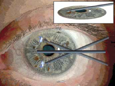
As the new study shows, using a microincision aspiration cutter is a third, minimally invasive option that yields excellent biopsy results while minimizing some of the risks associated with the other two methods.

microincision aspiration cutter

Microincision aspiration cutter-assisted iris biopsy is a variation of the Finger iridectomy technique developed by Dr. Finger. It uses a single, small corneal incision, which is safer than the larger incision required by wide-incision iridectomy. Dr. Finger explained the significance of this advancement in biopsy techniques for patients:

“Conducting a biopsy through a tiny incision like this means you don’t have to have stitches. Think of it like the developments in back or abdomen surgery over the past few decades. Surgeons used to have to really open the patient up, which often meant more pain and a longer post-op recovery. Then they developed microincisions to conduct surgery. This is the ophthalmic version of those surgical enhancements – a surgery upgrade, if you will.”

In this study, seven patients suspected to have diffuse iris melanoma underwent microincision aspiration cutter-assisted iris biopsy. Five of the patients were ultimately diagnosed with melanoma. None of the patients lost visual acuity, developed biopsy-related glaucoma, nor experienced hypema (bleeding in the eye). In fact, 75% of the patients showed reduced intraocular pressure after the procedure.

Microincision aspiration cutter-assisted iris biopsy can provide doctors with a safer alternative to the other two commonly used methods. It allows effective biopsies with a small incision, rapid rehabilitation and potentially fewer complications.


UK Patient’s Story Shows Importance of Routine Eye Exams for Cancer Detection

Ralph McMurray of Suffolk, England, was 66 years old when he had his first ever eye examination.

It revealed he had eye cancer.

West Suffolk Hospital

According to a local news report, the examining optometrist found a detached retina and referred McMurray to the West Suffolk Hospital for further tests, which revealed a large tumor in his eye – an ocular melanoma. Physicians at St. Bartholomew’s Hospital in London removed McMurray’s eye just weeks later due to the size of the tumor.

“I am so glad I went for that eye test as the cancer would have only got worse and I may not even be here now,” said McMurray. “I am now determined to do as much as I can to raise awareness of the importance of having regular eye tests… In cases such as mine early diagnosis is crucial and the tumor would have been spotted much sooner had I been having regular eye examinations.”

McMurray’s experience underscores the importance of routine eye exams and the importance of all eye care professionals learning Dr. Fingers’ MOST mnemonic (Melanoma = Orange pigment, Subretinal Fluid, Thickness of 2 or more millimeters) to effectively smaller intraocular melanomas.

The earlier a tumor is detected, the more likely vision- and life-saving treatments can be employed.

McMurray was fortunate the cancer was found, even though it had already advanced. Prompt treatment after diagnosis likely saved his life.

For UK nationals considering a second opinion or treatment in the United States, The New York Eye Cancer Center offers special programs to assist international patients. Click here to learn more.


ICO/ECF Join Forces to Treat Childhood Eye Cancer in Underserved Countries

Proper Training Expected to Substantially Reduce Mortality Rates 

The Eye Cancer Foundation is pleased to announce a partnership with the International Council of Ophthalmology (ICO) to promote and conduct eye cancer fellowship education for physicians from unserved countries.

The Eye Cancer Foundation and International Council of Ophthalmology

The ICO represents and serves professional associations of ophthalmologists from around the world. The organization’s mission is to “work with ophthalmologic societies and others to enhance ophthalmic education and improve access to the highest quality eye care in order to preserve and restore vision for the people of the world.”

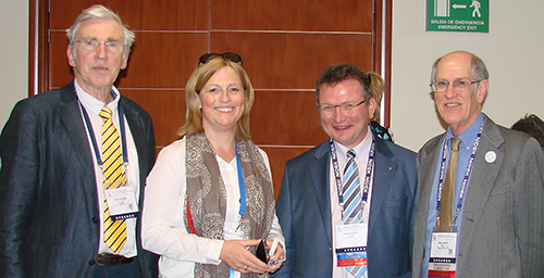
The ICO Fellowship program leadership team
Left to right: Peter Gabel, MD (former ICO Fellowships Director), Cordula Gabel-Obermaier, MD (ICO Fellowships Executive), Berthold Seitz, MD (ICO Director for Fellowships), and Bill Felch (ICO CEO).

This new partnership will expand and enhance The Eye Cancer Foundation’s existing fellowship programs. It will focus specifically on addressing the need to reduce world-wide mortality related to retinoblastoma by ensuring that more patients have ready access to a trained eye cancer specialist in their region.

Untreated childhood retinoblastoma

Retinoblastoma is the most common eye cancer in children. It typically develops by the age of five. Retinoblastoma affects approximately 8,200 children each year. The incidence rate is somewhat higher in developing countries, where most of the children succumb to metastatic retinoblastoma. In contrast, there exists a better than 96% survival rate in developed countries like the USA.

Early diagnosis and prompt treatment will save lives. In countries without eye cancer specialists, children with retinoblastoma typically go undiagnosed and untreated until it is too late. Local general ophthalmologists are not familiar with modern eye cancer medicine. Training doctors to diagnose and treat retinoblastoma in these areas will save lives. The Eye Cancer Foundation and the International Council of Ophthalmology has set a goal of saving 1,000 children by 2020.

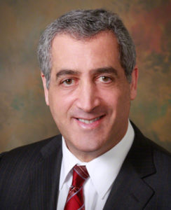
Paul T. Finger, MD
Dr. Paul T. Finger, Chairman, The Eye Cancer Foundation

The Eye Cancer Foundation currently funds 25 half-year retinoblastoma fellowships in ophthalmic oncology for candidates from unserved or underserved countries. The new partnership with the ICO will help expand the scope and reach of this fellowship program.

“The Eye Cancer Foundation is looking forward to a long and productive relationship with the International Council of Ophthalmology,” says our Chairman Dr. Paul Finger


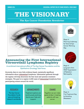
The Visionary Newsletter – Summer 2016 Edition

The following was originally published by The Eye Cancer Foundation.

The Visionary Newsletter - Summer 2016 EditionThe Summer 2016 edition of The Visionary is now available! The Eye Cancer Foundation publishes The Visionary free-of-charge to keep you informed about the latest news, research, and global efforts focused on improving eye cancer treatment, diagnosis, and cure.

In this edition, you will find articles about:

  • The First International Vitreoretinal Lymphoma Registry
  • The Second Eye Cancer Working Day in 2017
  • A Massive International Study Yielding Key Insights into Preventing Metastasis
  • And More

The Eye Cancer Foundation can send The Visionary directly to your inbox. If you’d like to receive a personal email notification when new editions of The Visionary are released, please follow this link to the eyecancercure.com website. Your information will not be shared with third parties.


Patient Stories

"Very well treated by Dr. Finger. He explained everything I needed to know about my issue with detail and attention, putting me at ease and giving me confidence to handle this problem for the rest of my life.”
N.N.

Learn More About Our Patient Care

REQUEST AN APPOINTMENT

Go to Appointment Form

CONNECT AND SHARE