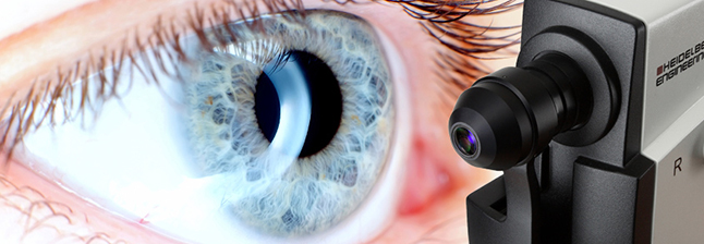
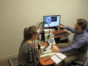
The New York Eye Cancer Center is thrilled to announce it is the first clinic in the United States to use the state-of-art OCT2 imaging machine from Heidelberg Engineering. The machine was publicly released earlier this year and installed at NYECC the week before Christmas. It reflects Dr. Finger’s continuing commitment to provide cutting-edge, next-generation eye cancer care. Dr. Finger insists on the best for his patients, and the OCT2 will greatly improve the accuracy of our diagnoses even further.

Dr. Finger pioneered the use of OCT (Optical Coherence Tomography) for radiation retinopathy. The OCT technique creates 3D images of the retina and is one of the best tools available for the diagnosis of eye cancer. The OCT2 represents a huge step forward with this technology, allowing Dr. Finger and his staff to make even more precise diagnoses.
OCT is similar to an ultrasound, but uses light instead sound waves. As a result, it offers much clearer images that are superior to traditional MRIs or ultrasounds. The OCT imaging technique uses light to capture 3-D images from within tissue, creating high-definition pictures at resolutions comparable to a low-power microscope. Using the OCT2, Dr. Finger will be able to see within, around, and even behind intraocular tumors. For his patients, this means better diagnoses, treatments, and ultimately better outcomes.
The OCT2 features significant advances over the older technology. Its extremely fast scanning speed improves scan placement and limits interference, known as artifact potentials. This faster scan also includes rapid data acquisition, which allows the machine to record even more data than before and provide:
- 3D visualizations
- High-resolution images
- Ultra-high density scans
Looking at blood flow within the internal structures of the eye – angiography – is vital to accurately diagnosing eye cancer. The OCT2 is capable of recording high-speed angiography and can actually create movies of blood flow dynamics. It’s also all ready to be upgraded to the emerging technology of dye-less angiogram imaging.
Ultimately, the OCT2 will allow Dr. Finger to view more of the eye at one time. It features Full Depth Imaging (FDI), allowing visualization of vitreous (the clear gel that fills the space between the lens and the retina) to choroid (the vascular layer of the eye containing connective tissue that lies between the retina and the sclera) in one single image.

On top of this, a new wide-field capability allows Dr. Finger to see the anterior retina. It can tune in different wavelengths of light to image at different depths within the retina itself. The ultra-wide field of vision offered by the OCT2 allows for more of the retina and tumor to be seen within a single image.
For the time being, the NYECC is the only clinic in the United States where patients can have their eyes imaged with this cutting-edge machine. Until now, it has only been in use at various research sites in the US.
Not only will Dr. Finger have one of the best eye cancer diagnostic tools at his fingertips, but the machine’s faster scanning also greatly improves the patient’s experience at NYECC. Patients will spend less time staring into a machine and more time discussing their case and the best course of treatment with Dr. Finger. Eventually the OCT2 will be upgraded to dye-less angiogram imaging, meaning patients won’t need to receive dye injections.
The acquisition of the OCT2 machine continues NYECC’s commitment to being the next generation of eye cancer care. By combining the most technologically advanced diagnostic tools with pioneering treatments, Dr. Finger offers his patients the most advanced care available.


