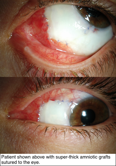
Rebuilding the Eye Using Amniotic Membrane Grafts
Byline: Published in The American Journal of Ophthalmology, 2019;198:45-53
Since 1940, a single, thin layer of amniotic membrane graft (AMG) has often been used for repairing the cornea and conjunctiva. However, Dr. Finger says:
“our research shows that super-thick AMG (ST-AMG), up to ten times thicker than the prior AMG, is more effective for reconstruction of the eye’s surface.”
 Research supported by The Eye Cancer Foundation has proved greater efficacy of this new technique in recreating the outer surface of the eye and inner surface of the eyelids. As published in the American Journal of Ophthalmology on November 2018, tumors of the conjunctiva and eyelids were surgically removed, then amniotic membranes from donor human placentas were sewn into the defects to recreate a normal ocular and inner eyelid surface.
Thus, amnion can provide a foundational platform for new cells to grow and flourish. In this case series, super thick amniotic membrane grafts (AMGs) were found to facilitate the healing of wounds.
How exactly does graft thickness affect the success of treatment? Well, the greater thickness means it is more easily sewn into the affected area, and
also helps the grafts to remain several weeks after placement. Thicker grafts are less likely to tear, rupture, or dissolve during the postoperative period. Most importantly, following treatment with ST-AMG, every single patient retained their sight and found their wounds successfully healed.
Super-thick amniotic membrane grafts have proven benefits to their thinner counterpart, and perhaps its versatility hints at potential for greater medical applications in the near future.
To keep up-to-date on the latest in Eye Cancer News, bookmark our website or follow us on facebook!
Click here to read the full research paper
Funding/Support for this study was provided by The Eye Cancer Foundation, Inc.
Click here to donate to further advancements in eye cancer research.


