With a minuscule incidence of less than 0.005%, a myxoma is a staggeringly rare condition. Defined as noncancerous tumors of our connective tissue, myxomas present similarly to other, slightly more common conditions such as conjunctival lymphoma, lymphangioma, ocular surface squamous neoplasia (OSSN), or amelanotic melanoma. Consequently, they are usually misdiagnosed, or, at the very least, are difficult to diagnose.
Unique Ultrasound Findings!
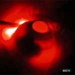
Very limited literature exists describing cases and interventions of myxoma. In an effort to offset this shortage of research, The Eye Cancer Foundation sponsored a publication describing a myxoma case with unique ultrasonographic findings.
Other tumors of the conjunctiva often appear as a single, solid mass. Myxomas in particular present as smooth, yellow-pink masses on the eyeball, ranging in size from 4 mm to 20 mm. In this particular case under study, the patient’s myxoma showed scattered cells rather than a uniform image. So, while they are similar in many ways to other tumors, myxomas can have unique features that separate them from the others.
The Verdict…
Dr. Finger, Chairman of the ECF and Chief Researcher in this case study, concludes that:
“Though conjunctival myxomas can masquerade as various other conditions, high-frequency ultrasound proves myxomas have a distinct vascular pattern and no evidence of intraocular tumor invasion.”
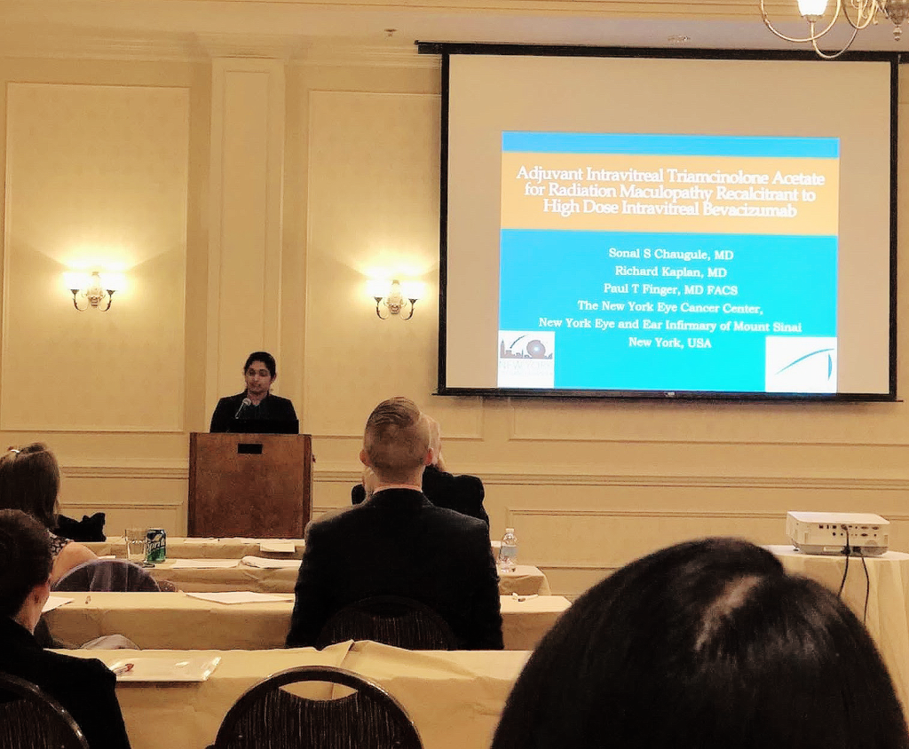
The New York Eye Cancer Center and the Eye Cancer Foundation were quite actively represented at the 2017 American Association of Ophthalmic Oncologists and Pathologists (AAOOP) Annual Meeting. The meeting was held on Friday, November 10, 2017 at the Hampton Inn & Suites Convention Center, located in the vibrant city of New Orleans, Louisiana, and was attended by Dr. Paul Finger as well as notable ECF-ICO Fellowship alumni, Dr. Sonal Chaugule, Dr. Ekatrina Semenova, and Dr. Abhilasha Maheshwari.
Now, what are ITA, RM, and Bevacizumab? Often, patients undergoing eye plaque radiation in order to treat their cancerous tumor can be subject to vision-impairing radiation side-effects, or radiation maculopathy (RM), as a result of treatment. Intravitreal anti-VEGF therapy (which is otherwise commonly used to treat macular degeneration) such as Bevacizumab (Avastin), Lucentis, and Eyelea, are used to prolong the effects of radiation maculopathy. Itravitreal triamcinolone acetate (ITA) is a steroid used in conjunction with this anti-VEGF therapy to treat swelling that occurs in the affected eye, called macular edema.
The paper aims to evaluate the effects of using ITA for the treatment of RM in patients with choroidal melanoma after plaque radiotherapy. Eight choroidal melanoma patients undergoing this treatment were studied, having ITA treatment at 4-16 week intervals in addition to continued injections of Avastin. Results found that after starting ITA, vision was stable or improved for patients, leading to the conclusion that ITA can be used as a supplement to decrease macular edema (swelling) and preserve vision in choroidal melanoma patients with RM.
Dr. Abhilasha Maheshwari had separately presented ECF-supported research — a 12-year study evaluating patients with slotted, low energy photon eye plaque radiation therapy. The purpose? To measure the efficacy of this treatment for eye cancer patients, especially those who have tumors located near, touching, or surrounding the optic disc (a critical area that allows for vision) were treated. Forty six patients of these eye cancer patients were treated with eye plaque radiation therapy, using seeds of the chemical isotope Palladium-103 to radiate the affected eye. Over the next 12 years, these patients were monitored for any changes to tumor thickness, visual acuity, and whether or not the cancer had reoccured or metastasized. Results found that the local control rate (i.e, total tumor destruction) was 95.6%, and lead to the conclusion that Slotted Eye Plaque Radiation Therapy is indeed an efficient method of treatment for eye cancer patients. To read the paper, published in the American Journal of Ophthalmology, click here.
But the AAO updates do not end here! Stay tuned for upcoming information on even more presentations at AAO 2017 by ECF alumni.
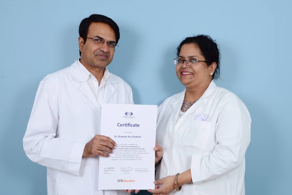
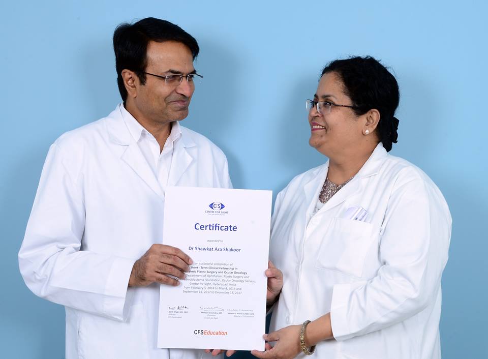
The Eye Cancer Foundation 2020 Campaigncontinues to make tremendous strides in the advancement of eye cancer care through the exciting completion of Dr. Milly Shakoor’s 6-month fellowship in retinoblastoma training. This news arrives unitedly with the announcement of another ECF Fellow’s completed training, Dr. Veronica Molleda, from Bolivia. With every fellowship thus offered and completed, The Eye Cancer Foundation and its supporters come closer to fully realizing the goal of training 20 specialists in 20 countries to treat childhood eye cancer.
Eye Cancer Foundation fellowships offer doctors to be trained in the specialized treatment of retinoblastoma, training that they cannot otherwise receive in their home country. These ECF fellowships, partnered with the International Council of Ophthalmology (ICO), are available to candidates from unserved or underserved countries. After doctors complete their six months of training, they agree to return to their home country to start or participate in eye cancer treatment for the unserved.
But what is retinoblastoma? Retinoblastoma is the most common eye cancer in children and affects approximately 8,200 children each year. In developed countries like the United Sates,the survival rate reaches beyond an astounding 96%, with early diagnosis and treatment being key to saving patients’ lives and sight. However, the incidence rate is higher in developing countries, where most of the children succumb to metastatic retinoblastoma. In areas where children and families have no means of traveling to treatment centers far away from them, these afflicted children often endure their untreated disease untreated, which eventually leads to death. Because no child or family should have to suffer these losses, especially due to inability to simply reach a treatment center, the ECF has launched the 2020 Campaign.
Dr. Milly Shakoor comes from Dhaka, the capital of the highly densely-populated country of Bangladesh, where availability of retinoblastoma care is certainly low. She trained at The Centre for Sight in Hyderabad, India with the renown Director of Medical Services, Dr. Santosh G. Honavar (pictured above), who specializes both in oculoplasty and ocular oncology. Since her return to Dhaka, she has been met with several retinoblastoma cases and continues her treatment of them.
As always, The Eye Cancer Foundation these fellowships could not have been completed without the support of readers and donors — and so, the ECF thanks you for helping to provide hope for eye cancer patients around the world. To our audiences, we hope that you will continue to support these projects through your continued readership, word of mouth, and well wishes!
As you may have read in our blog the week prior, The New York Eye Cancer Center is pleased to announce that we, with the support of The Eye Cancer Foundation, are hosting periodic group therapy sessions for our patients. Life after diagnosis and treatment of an ocular melanoma can cause stress and anxiety. Although it’s normal to feel this way, many people do find that having a solid support system is crucial in finding their bearings and coming to terms with “a new normal”, both of which are important in overall quality of life.
Friends and family can be excellent support systems, but there can also be a benefit to sharing your feelings with other patients who have had very similar experiences. The New York Eye Cancer Center Support Group is seeking to provide you with this emotional outlet. Our support group is facilitated by wonderful licensed clinical social worker, Karen Campbell.
You can find Karen’s short biography below:
“I am a Licensed Clinical Social Worker (LCSW) and have been practicing in the field for 24 years. In addition to my private psychotherapy practice, most recently, I was in the Director of Vision Rehabilitation Services at the Lighthouse Guild. I started up the Social Work Department at Lighthouse International in 2010 and, as part of that, developed and faciliated the Department’s Support Group program. I also have a background in medical social work, including oncology, having worked at NYU/Langone. I have found support groups to be a valuable way for people to manage their medical challenges and address issues such as family adjustment, depression, anxiety and loss. Although I work primarily with individuals and couples in my private practice, I really enjoy working with groups!”
Karen brings to the practice her years of experience in counseling patients with both cancer and vision loss, making her a perfect fit for the NYECC family.
You can meet her and other patients at our next group therapy session at the NYECC on Friday, October 13, 2017 at 1:30 PM, Eastern Standard Time.
We hope to see you there!
And to stay updated on all upcoming sessions, please keep our website, eyecancer.com in your bookmarks!
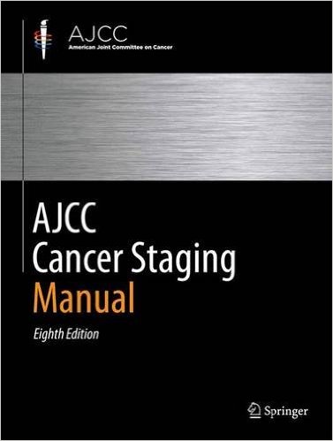
“Sharing a common scientific language (staging systems) allow us to communicate our ideas and enable progress” writes Dr. Paul T. Finger in Foundational Elements for Collaboration in Ophthalmic Oncology; a recent editorial published for the American Academy of Ophthalmology.
Communication is vital in nearly all areas of life, and the medical field makes no exception. In order for doctors to compare their results, they must describe the stage (size and distribution) of the cancer they are treating. This is the only way oncologists can effectively discuss and coordinate the care for patients around the world. And towards this goal, Dr. Finger as Chair of the Ophthalmic Oncology Task Force for The American Joint Committee on Cancer has worked over 12 years to create, write, and publish three editions of The AJCC Cancer Staging Manual and most recently its sister equivalent for the UICC.
As Dr. Finger explains, the AJCC staging system was made from the collective effort of 10 subcommittees, composed of more than 50 eye cancer specialists across the world. These doctors came together to develop a clinically useful textbook describing, with rigorous detail, the methodology of classifying eye cancers. In fact, working together, these systems represent the greatest consensus work yet created by the eye cancer specialty.
You may have heard of a tumor classified as “stage 2” or “stage 4”, benign or malignant, and so on, but how do doctors come to this conclusion? Staging a tumor relies on rules on how to measure and locate tumors in the eye and/or thought the body. By gathering a large profile of data from tumor patients, the AJCC team has also analyzed how tumor size or failure of initial treatment can be used to predict the risk for metastasis.
With this common staging system shared between doctors from The United States to Indonesia, patient care is streamlined and made more effective. The AJCC’s wide pool of data allows for a more precise system, which is tremendously useful for eye cancer specialists otherwise unable to access international resources. This is the immediate effect of doctors adopting this system, but the long term effects are equally as influential.
And to stay updated on all Eye Cancer Foundation news, as well as information on the latest eye cancer research, please keep eyecancer.com in your bookmarks!
This eye and vision sparing treatment utilizes a metallic plaque, sometime called a “radiation implant” or “radioactive source.” The doctor surgically implants the plaque on the wall of the eye, covering the base of the intraocular tumor. The implant remains in place for five to seven days, delivering a highly concentrated radiation dose to the tumor. The plaque’s location on the eye means surrounding healthy tissues get relatively less radiation exposure.
Once the plaque is in place, The New York Eye Cancer Center patient spends the rest of the treatment period at home or at a hotel. After the prescribed amount of time, the patient returns to the hospital to have the plaque safely removed.
The following video explains the procedure, what to expect, and the safety measures that must be followed during the course of treatment.
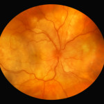
A small choroidal breast metastasis, the larger tumor was in the other eye.
Malignant tumors from other parts of the body can spread in and around the eye. These tumors may never be discovered unless they affect vision, are visible to the patient, or push the eye forward. The most common location for ocular metastasis, in the vascular layer called the choroid (choroidal metastasis) within the eye.
Cancer metastasis that appear in and around the eye are usually from a breast cancer (in women) and lung cancer (in men). Other less common sites of origin include the prostate, the kidney, the thyroid, and gastrointestinal tract. Blood cell cancer (lymphoma and leukemia) can also metastasize to the eye and orbit. Once a patient is diagnosed with choroidal metastasis, we try to find where it came from. In 18% of patients, we do not find the source of choroidal metastasis. In these cases, we may have to biopsy the ocular tumor and look at its cell-type.
A small choroidal breast metastasis, the larger tumor was in the other eye.
Symptoms
Most patients with choroidal metastasis have no symptoms. If the metastasis is on the eye or eyelids, it may be visible. If located behind the eye (in the orbit), the metastasis can push the eyeball out or to the side. If within the eye (the most common), choroidal metastasis patients can see flashing lights, floating spots or distortion of their vision. Patients with a history of cancer are at greatest risk and should have periodic eye examinations.
Diagnosis
Most patients with metastasis have either a known primary cancer and/or metastatic tumors in other parts of their body. A careful medical history can uncover the signs or symptoms of these other cancers. If an eye cancer specialist suspects ocular metastasis, both eyes and orbits should be examined because ocular metastases can be both bilateral and/or multifocal.
Choroidal metastasis is usually non-pigmented (except metastatic melanomas), and has typical ultrasound and angiographic patterns. Choroidal metastasis is usually poorly circumscribed and can cause retinal detachments. They may have spicules of pigment on their surface. Unlike primary choroidal melanoma, they can grow quickly (weeks) and may require prompt treatment.
The patient with metastasis to the eye should also be examined by a medical oncologist. A complete metastatic survey should be performed to “stage” the patient (to see if there are other tumors within the body). Specifically, computed radiographic imaging of the brain and lung should be performed due to a high concurrent incidence of intracranial and pulmonary metastases. Your doctor may suggest a total body PET/CT with fusion.
Treatments
The care of patients with metastasis to the eye typically involves cooperation between the eye cancer specialist, medical oncologist, and radiation therapist. Though chemotherapy can be used in many cases of orbital and choroidal metastasis, radiation therapy is usually a more definitive treatment. If the metastatic tumor has not destroyed the center of the retina, early treatment offers the best hope for preserving vision. Almost all patients with choroidal metastasis can be treated with external beam radiation. That is, surgery is rarely needed as treatment for choroidal metastasis. Orbital and adnexal ocular metastasis are typically biopsied prior to treatment.
After irradiation, a regressed choroidal metastasis displays spicular hypertrophy of the retinal pigment epithelium.
Most patients who develop posterior choroidal metastasis can either be closely monitored for tumor growth, followed for response to chemotherapy, or treated with external beam irradiation. Radioactive plaque radiotherapy is rarely needed. If chemotherapy is not an option, prompt external beam irradiation (typically 30-40 Gy), may offer the best chance for preservation of vision.
In those rare cases where the metastasis spreads to the iris, patients can develop severe glaucoma and may lose their eye. Thankfully, anterior segment (iris) metastases are rare. Since most cancers do not spread to the iris, most patients with intraocular, choroidal and orbital metastasis respond well to treatment and keep their vision.
"Very well treated by Dr. Finger. He explained everything I needed to know about my issue with detail and attention, putting me at ease and giving me confidence to handle this problem for the rest of my life.”
– N.N.