2020 Fellowship

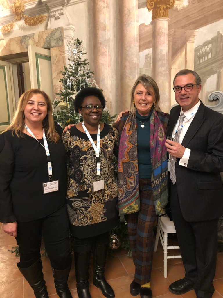
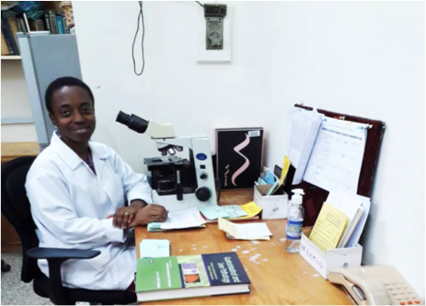
Ghana is another country claimed by the Eye Cancer Foundation! Recently, Dr. Akosua Boateng finished her training sponsored by The Eye Cancer Foundation (http://eyecancercure.com). She notes, “I have always been interested in ocular pathology, but had never truly had the opportunity to nurture the interest due to a lack of resources in my home country.” However, she was awarded the opportunity to train in ophthalmic pathology with an emphasis on retinoblastoma at the Beijing Children’s Hospital due to a joint effort of Dr. Paul Finger of The Eye Cancer Foundation and Dr. Brenda Gallie of the University Health Network, Princess Margaret Cancer Center of Toronto. 


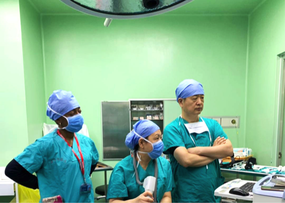
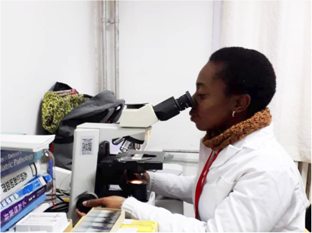
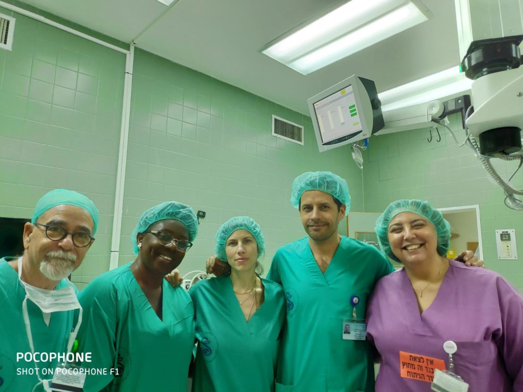
Dr. Boateng was determined to learn about the clinical presentation, examination, and treatment for many ocular cancers as well as be able to recognize their pathology on slides under the microscope.  Her training 

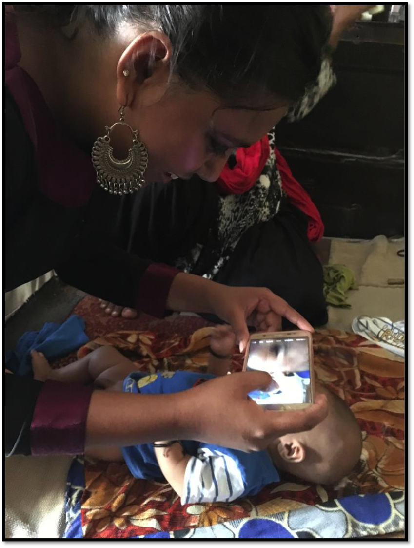
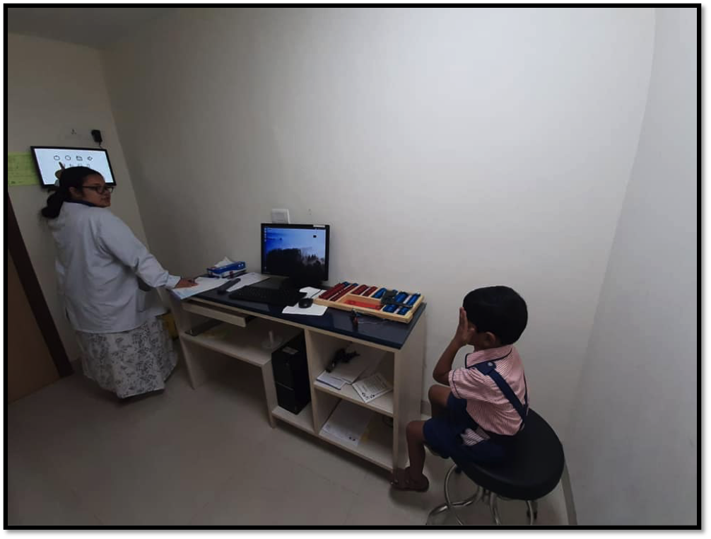
was split between a clinic with Dr. Jeffrey Zhang, retinoblastoma care with Dr. John Zhao and the pathology department with Dr. Nan Zhang. During clinic days, Dr. Boateng learned about many eye diseases including myopia, congenital anophthalmia and others. For the first time, she learned how to perform intraocular photography with the Retcam Imaging System. For retinoblastoma care, Dr. Boateng was able to examine the fundus of both old, treated  and new cases. She took part in discussions of the children’s treatment options. She spent time in the operating theatre shadowing surgeries, such as laser treatment, chemotherapy and enucleation. She learned to differentiate between retinoblastoma and Coat’s Disease. In the pathology department, first she learned more about the anatomy of the eye, identified histologic high-risk factors for retinoblastoma, and how to stage cases according to the AJCC 8th Edition Manual. She says it was a “great learning experience.”. 


Unfortunately, due to the Covid-19 pandemic, Dr. Boateng was forced to abruptly end her training months prior to completion. Thus, she would like to return to learn more about adult ocular pathology as it would be helpful back in Ghana. However, when Dr. Boateng returned to her parent hospital in Ghana, she feels so much more confident in dealing with ocular pathology and retinoblastoma. She is grateful to Drs. Finger and Gallie as well as The Eye Cancer Foundation for allowing her intensive training to happen! 


3 – Year Journey – Raising Awareness for Retinoblastoma

Retinoblastoma is the most common intraocular cancer in children. One out of 15,000 children suffer from this malignancy. Typically, parents notice that one or both pupils turn white and their child has difficulty seeing.  In 2017, a university in New Delhi, India initiated a long-term project to detect that white eye reflex in children. It was spearheaded by Nirmala Muralidhar, the project coordinator of Janki Devi Memorial College, under the direction of Dr. Vikas Menon, their consultant ocular oncologist. What began as a student faculty project, in November 2017, it transformed into a larger initiative where a Non Governmental Organization (NGO), ophthalmologists, ocular oncologists from specialty eye cancer centers collaborated to create awareness for early detection of retinoblastoma cancer.  


The long-term goals of this project included sensitizing families to the appearance of the white eye reflex, stirring awareness amongst doctors about retinoblastoma and prompting parents to be aware of their children’s eye health. Using photography as a tool for early detection, they created awareness of children with special needs who suffer from retinoblastoma. Though these objectives were slowly accomplished over 3 years, there were many activities, learning opportunities, and research studies presented to educate the public. The Strides for Retinoblastoma was most profound and heartfelt, as an RB survivor led a walk with over 200 people during World Cancer Day on February 4, 2018. 


While many research studies are able to spread awareness of retinoblastoma within the healthcare professional world, this initiative is unique as its audience was varied. It included parents, children and healthcare workers. All were involved in this coordinated campaign to save both vision and life.. The Eye Cancer Foundation continues to be determined to create awareness of retinoblastoma, to supply trained eye cancer specialists to underserved communities and is more than proud to announce the success of the JDMC awareness campaign. 


For more information contact  Dr. Puneet Jain at pjain@eyecancer.com

Retinoblastoma Screening in Babies

Public Awareness Campaign 

Eye Checkups


Message From The Eye Cancer Foundation

Did you know…


…that in addition to the training of eye doctors from around the world, The Eye Cancer Foundation needs to equip them with essential basic eye cancer care equipment. Especially during COVID-19, eye cancer specialists in underserved countries are having difficulty treating their patients with basic equipment designed to help pinpoint the disease. Without the proper equipment, these patients will lose their vision, partially or completely or required eye removal “enucleation.”  Help us equip these eye cancer specialists with essential items for their practices. Thankfully, Amazon makes this very easy.  Just go to the ECF Amazon page and select an item for as little as $6.99.  To get to the right Amazon page, just click here. Send it as a gift or write our Foundation’s Director of Development (henry@eyecancercure.com) to let us know what you have donated and that it will be coming. You can select from any of the equipment listed and donate as many as you want. Be assured, it will be used for eye cancer patients around the world. No matter what you donate, you will empower an eye doctor somewhere else in the world to save sight and save lives. Thank you, in advance, for helping us with this important work.


ECF Fellow, Dr. Ankit Tomar, Presents Research at the AAO & AAOOP!

In 2019, The Eye Cancer Foundation’s Fellow, Dr. Ankit Tomar, successfully presented two research projects at medical conferences in San Francisco, USA. To eye cancer specialists and ophthalmic pathologists at the AAOOP and American Academy of Ophthalmology Annual Meetings, Dr Tomar presented on Ciliary Body Leiomyoma: Clinicopathologic Correlation in 3 Cases. In his series, Dr. Tomar shows how ciliary body leiomyomas can be indistinguishable  from ciliary body melanoma. Only special pathology testing of tumor tissue samples are needed to separate these clinical dopplegangers.  


Dr. Tomar also presented on Dr. Finger’s well-known innovation, the ophthalmic palladium-103 plaque brachytherapy. In this study, he explored the safety and tolerability of this method for patients with multifocal iris melanoma. This study was entitled Palladium-103 Plaque Therapy for Multifocal Iris Melanoma: Radiation of the Entire Anterior Segment. Drs. Finger and Tomar discovered this method was highly effective for  this ocular cancer treatment since 1990! In this series, plaque radiation therapy achieved 100% local control and eye retention! This is remarkable because eyes with diffuse iris melanoma are commonly removed (enucleated).


The Eye Cancer Foundation is proud to support research that improves methods of diagnosis and treatment for eye cancer. Our fellows earn grants to present ECF research and learn about cutting edge technology and techniques. By spreading knowledge of ECF studies and findings, The Eye Cancer Foundation and its fellows like Dr. Ankit Tomar spread hope for those who would otherwise lose both sight and life. 

Dr. Finger (left) and Dr. Tomar (right)

Dr. Tomar’s presentation poster on Ciliary Body Leiomyoma: Clinicopathologic Correlation in 3 Cases.

Dr. Tomar’s presentation poster on Palladium-103 Plaque Therapy for Multifocal Iris Melanoma: Radiation of the Entire Anterior Segment.


AJCC Celebrates 60th Anniversary

This year, 2019, is the 60th anniversary of the founding of the American Joint Committee on Cancer, the AJCC. This committee was founded in 1959 by leaders of the American College of Surgeons in order to support patients by creating a staging system for cancer. The founders had the foresight as well as the influence to develop a cancer classification system based on the tumor, nodal status, and metastatic findings (TNM). What is interesting is that these features were first used by a French surgeon in the 1940s to classify breast cancer! They later became the standard international language for the staging system locked in place today by the AJCC for the needs of US physicians and hospitals. 

As the decades went by, the AJCC collaborated with the Union for International Cancer Control (UICC) to create a standardized cancer language for the world. Through the hard work of numerous physicians dedicated to improving cancer mortality, a worldwide TNM system was achieved in 1987. Since 1997, AJCC has published periodic improvements to the TNM system. 

In 2005, our very own Dr. Finger was selected to join the American Joint Committee on Cancer: Section on Ophthalmic Oncology! Then, 4 years later he was elected Chair, for Ophthalmic Oncology. Dr. Finger has participated in the AJCC for over 14 years. His work with the AJCC includes developing the AJCC Ophthalmic Oncology Task Force of over 50 eye cancer specialists from 13 countries on 6 continents. Dr. Finger put together specialty committees to develop a useful classifications to standardize how doctors describe their patients tumors and patterns of metastatic disease. For example, in 2014 the American Brachytherapy Society’s Ophthalmic Oncology Task Force adopted the AJCC staging rules for intraocular melanoma! 

Dr. Fingers’ hard work and dedication is further epitomized by his creation of The Eye Cancer Foundation, a community that aims to provide support to patients with ocular cancers, promote the education of the diagnosis and treatment of eye cancers, and train eye cancer specialists around the world. Numerous physicians from underserved and unserved countries have been trained under the sponsorship of the ECF and gone on to use their newfound knowledge to benefit patients in their home countries. 

The ECF will never stop saving lives and saving sight.

Click here to make a donation to the foundation and save the life of a child suffering from retinoblastoma!


Dr. Ann Musika Completes ECF Fellowship

From Uganda to Israel, and Back Home Again

Hailing from Kampala, Uganda, Dr. Ann Musika has completed her 6-month ophthalmic oncology fellowship training sponsored by the Eye Cancer Foundation.

From Left: Dr. Ann Musika, Dr. Alezam, Dr. Didi Fabian, and Dr. Vicktoria Vishnevska-Dai

For the past six months, Dr. Musika has trained at Sheba Medical Centre at the Goldschleger Eye Institute under supervision of the director of the ocular oncology unit, Dr. Vicktoria Dai. Her training included all aspects of eye cancer care––from diagnosis, to treatment, to intravitreal injections for radiation retinopathy. Additionally, she took part in various research studies at Goldschleger’s ophthalmology department.

The Eye Cancer Foundation places an emphasis world-class training as well as practical and applicable training. So while Dr. Musika learned all aspects of eye cancer treatment practiced at Goldschleger’s, she specifically focused on treatments and techniques that would be just as readily available to her in Kampala.

Dr. Ann Musika (second from the left) alongside Senior ocular oncologists (Presenters) with after the ocular oncology training at the University of Sienna Italy on December 6th-7th, 2018

“It is with great pleasure that I express my sincere gratitude

to you for the great support that you offered me…all this wouldn’t have been possible without your generous support. The knowledge and skills acquired from this training I hope to apply to improve the quality of care for our ocular oncology patients in my nation. I am forever grateful.”

Prior to Dr. Musika, there were no fellowship-trained eye cancer specialists in Uganda. Consequently, patients with eye cancer suffered not only from their disease, but also from a lack of specialized care. However, now upon the completion of her training, Dr. Musika intends to return to Uganda and provide her nation with much-needed specialty care.


To Learn More about the Eye Cancer Foundation, its mission, and fellowship opportunities, visit the website at www.eyecancercure.com.


Day 3: Live From the ISOO 2019!

Day 3 at the ISOO: Melanoma, Other Intraocular Cancers, and Basic Retinoblastoma Science

Every two years, eye cancer specialists worldwide convene at the International Society of Ocular Oncology Conference in order to review latest research and encourage international collaboration within the field of ophthalmic oncology.

For details on the ISOO Biennial Conference, read more here.


Dr. Puneet Jain presenting his iris varix poster

Dr. Puneet Jain returned to give us some insight on iris varix by presenting his paper Iris varix: 10-year experience with 28 eyes, published alongside Dr. Paul Finger during his Eye Cancer Foundation fellowship. The study began with the intention to more clearly define iris varix, consistent characteristics, imaging findings, and note any consistent change that might occur with observation. The results showed that iris varix is “primarily located in the inferotemporal quadrant, not associated with dysmorphic pupillary findings, progression, secondary glaucoma, or malignancy.” Essentially, “iris varices were benign vasculopathies with no associated ocular or vision-related morbidity.”


Dr. Ekaterina Semenova’s poster presentation

Why are some cases naturally suppressed by the immune system? Dr. Ekaterina Semenova raised the question, noting five cases in which choroidal melanoma in the patient had spontaneously regressed in her paper Spontaneously Regressed and Apparently Dormant Choroidal Melanomas, also published alongside Dr. Finger. She opened up discussion to the audience, though consensus was that this would be an interesting topic for future research in order to determine the cause. Now that such cases have been documented, we have this data to look back on and, hopefully, make additions to.


Dr. Abhilasha Maheshwari (left) and Dr. Paul Finger (right) after another successful poster presentation!

Finger’s Slotted Plaques was recognized in Dr. Abhilasha Maheshwari presentation, A 12-Year Study of Slotted Eye Plaque Radiation for Choroidal Melanoma: Near, Touching, or Surrounding the Optic Nerve. Over a decade’s worth of data was analyzed and slotted plaques were found to be very effective and resulted in good patient outcomed for patients whose tumor lies close to the optic nerve. These plaques offer more proper positioning of the plaque, allowing radiation to the entire melanoma plus a 2-3 mm margin. Dr. Maheshwari concluded that even after 12 years, “slotted plaque radiation therapy resulted in high rates of local tumor control and vision and eye retention.”


That’s all for today’s ECF highlights! Day 4 will be fully dedicated to retinoblastoma. Stay tuned!


To help sponsor fellowships and fellow research, donate to The Eye Cancer Foundation at www.eyecancercure.com/donate.


Day 2: Live From the ISOO 2019!

Day 2 at the ISOO: Melanoma Day

Every two years, eye cancer specialists worldwide convene at the International Society of Ocular Oncology Conference in order to review latest research and encourage international collaboration within the field of ophthalmic oncology.

For details on the ISOO Biennial Conference, read more here.


How Can You Tell a Choroidal Melanoma is Regressing?

Dr. Abhilasha Maheshwari, ECF fellow

Dr. Abhilasha Maheshwari was the first Eye Cancer Foundation fellow to lecture today with her presentation on the paper Regression Patterns of Choroidal Melanoma after Plaque Brachytherapy, published alongside Dr. Paul T. Finger. In the two-minute rapid-fire session, Dr. Maheshwari filled us in on her data points from the study, concluding that there are various markers that show us when a choroidal melanoma is controlled after treatment with plaque brachytherapy. She provided us various markers, such as:

  • Dr. Finger and Dr. Maheshwari

    Increased tumor pigmentation.

  • Ultrasonography results:
    • maintenance of dome/mushroom shape
    • decreased tumor thickness
    • increased internal reflectivity
  • Fundus autofluorescence imaging showed a pattern of increased followed by decreased organge pigment lipofuscin
  • OCT showed decreased exudative subretinal fluid, and resolved drusenoid retinal pigment epithelial detachments (DRPED)
  • Flurocein angiography showed resolution of intrinsic tumor vascularity

Each of these characteristics serve as indicators of tumor regression. Dr. Maheshwari gave us one final take-away message: better initial local control means extremely less systemic spread, which overall means better patient outcomes.

Dr. Finger and Dr. Maheshwari in the “hot seats” up on stage – Every new session rotates a panel of doctors who have given lectures in order to ask and answer questions and foster discussion.

Interesting New Finding: One talk updated us on recent clinical trials for a drug that showed promise in laboratory studies for reversal of the effect of BAP1-related tumor progression. Perhaps we will hear more on this at the next ISOO!

Unique Case Report: Two, Simultaneous Primary Melanomas

Dr. Maheshwari returned for her second talk of the day, presenting results from Bilateral Simultaneous Primary Choroidal Melanomas: Treated with Palladium-103 Plaque Radiation, published along with Dr. Paul Finger. She elaborated on an interesting case who presented with separate, unrelated choroidal melanomas, one in each eye. After sequential treatment to maximize outcomes, the patient remains at 20/25 vision and shows great outcomes since their latest follow-up.


Unique Case Report: 7 Years After Treatment of a Primary Melanoma, A Second Primary is Found

Dr. Finger gave his first talk of the event to present a unique case of a patient who had been successfully treated for a stage T1 ocular melanoma. Seven years after treatment, however, she developed choroidal metastasis (this one, T3). Following intensive immunotherapy, the patient showed tumor controlled and marked decrease in liver metastasis. Upon follow-up, the patient still sees 20/25 and is maintained with anti-VEGF therapy.

With this talk, Dr. Finger wants to emphasize “the importance of consistent, long-term, post-treatment periodic ophthalmic and systemic surveillance for patients with choroidal melanoma.”

Dr. Finger Presents a Unique Case!

Identifying Genes Associated with Cancer Predisposition

Dr. Colleen Cebulla presented interesting findings in her presentation, Whole exome sequencing to identify candidate genes associated with hereditary predisposition to uveal melanomaShe covered various, possibly cancer-related genes other than the well-known, BAP1 mutation. At the beginning stages of research, the project has already analyzed 29 cases and identified a significantly large incidence of the genes SMARCE1,, PALB2, and MLH1. Particularly of interest are  PALB2, which has already been linked to breast, ovarian, and pancreatic cancer risk, and MLH1, which has been linked to Lynch syndrome and has been deemed responsible for multitudes of different tumors.

Dr. Cebulla is happy to welcome additions to the study and to be contacted if any centers are willing!


“SORTT” for Short: Study of Ophthalmic Radiation Therapy Toxicity 

Unfortunately unable to attend, Dr. Wolfgang Sauerwein requested Dr. Finger to present on Study of Ophthalmic Radiation Therapy Toxicity (SORTT): a prospective international survey. A major talk of the day, SORTT proposes “a prospective, multicenter, international data registry that collects structured information on toxicities after radiotherapy for eye cancer. This will offer the medical evidence needed for ophthalmic radiation side effect staging and modality selection.” This data may also help to create an AI to help physicians select the optimal radiation modality for each individual case.


Another jam-packed session of presentations, lectures, research findings, and plans for greater multicenter cooperation. As Day 2 comes to a close, Day 3 promises more on melanoma, other intraocular cancers, and the beginning of retinoblastoma discussions.


Day 1: Live From the ISOO 2019!

Day 1 at the ISOO: International Collaboration & Eye Cancer Care Worldwide

Every two years, eye cancer specialists worldwide convene at the International Society of Ocular Oncology Conference in order to review latest research and encourage international collaboration within the field of ophthalmic oncology.

For details on the ISOO Biennial Conference, read more here.

International Collaboration

The day began with a few talks instigating discussion on how to approach international, collaborative efforts, and how to improve on how we currently approach international cooperation. How do we get over international boundaries and laws restricting data-sharing, and varying laws surrounding research across countries? Much is yet to be done, though discussion is expected to continue as specialists from all over the world have convened at this conference.

Read on how Dr. Finger encourages international cooperation here.

What Do We Register and Why?

Featured ECF Fellow: Dr. Puneet Jain

Dr. Puneet Jain, on Registering Conjunctival Melanoma
Dr. Paul Finger (left) and ECF fellow Dr. Puneet Jain (right) immediately after his debut lecture.

Registries were a huge topic of discussion today. What is possible to be registered, what details should we include within the registry, how do avoid duplicate cases within separate registries, and how do we apply this to international cooperation? Among other points of discussion, however, it was unanimous that functional registries would assist greatly both with research and patient outcomes. The New York Eye Cancer Center’s recently graduated fellow, Dr. Puneet Jain, spoke particularly on the registering of conjunctival melanoma. His research with Dr. Finger evidenced the accuracy of the AJCC’s TMN classification for predicting metastasis.


Developing Worldwide Retinoblastoma Care

Dr. Brenda Gallie credits the Eye Cancer Foundation for research support.


Dr. Brenda Gallie discussed the development of retinoblastoma care worldwide. She noted the growing importance of genetic testing, stating a case in which laser treatment was sufficient enough to control the disease. She briefly mentioned project DepictRB as a way to display anonymized patient data, and offered the audience a demo of the program.

Conclusion

The day concluded with unanimous agreement that worldwide collaboration within the field of eye cancer needs to continue and needs to improve. With enhanced cooperation and worldwide registries, the

amount of shared data will assist greatly in advancement of research, diagnosis, and patient outcomes.

Tomorrow we cover ocular melanoma in great detail. Stay tuned, out fellows and Dr. Finger will be informing us with their expertise!

ISOO 2019 Day 1 Complete!

Rebuilding the Eye Using Amniotic Membrane Grafts

Byline: Published in The American Journal of Ophthalmology, 2019;198:45-53


Since 1940, a single, thin layer of amniotic membrane graft (AMG) has often been used for repairing the cornea and conjunctiva. However, Dr. Finger says:

“our research shows that super-thick AMG (ST-AMG), up to ten times thicker than the prior AMG, is more effective for reconstruction of the eye’s surface.”

Research supported by The Eye Cancer Foundation has proved greater efficacy of this new  technique in recreating the outer surface of the eye and inner surface of the eyelids. As published in the American Journal of Ophthalmology on November 2018, tumors of the conjunctiva and eyelids were surgically removed, then amniotic membranes from donor human placentas were sewn into the defects to recreate a normal ocular and inner eyelid surface. 

Thus, amnion can provide a foundational platform for new cells to grow and flourish. In this case series, super thick amniotic membrane grafts (AMGs) were found to facilitate the healing of wounds.

How exactly does graft thickness affect the success of treatment? Well, the greater thickness means it is more easily sewn into the affected area, and

 also helps the grafts to remain several weeks after placement. Thicker grafts are less likely to tear, rupture, or dissolve during the postoperative period. Most importantly, following treatment with ST-AMG, every single patient retained their sight and found their wounds successfully healed.

Super-thick amniotic membrane grafts have proven benefits to their thinner counterpart, and perhaps its versatility hints at potential for greater medical applications in the near future.


To keep up-to-date on the latest in Eye Cancer News, bookmark our website or follow us on facebook!

Click here to read the full research paper

Funding/Support for this study was provided by The Eye Cancer Foundation, Inc.

Click here to donate to further advancements in eye cancer research.


Patient Stories

"Very well treated by Dr. Finger. He explained everything I needed to know about my issue with detail and attention, putting me at ease and giving me confidence to handle this problem for the rest of my life.”
N.N.

Read More

REQUEST AN APPOINTMENT

Go to Appointment Form

CONNECT AND SHARE